Aurora 1510A Cardiac & Smooth Muscle Mechanical Testing System
| Origin | Canada |
|---|---|
| Manufacturer Type | Authorized Distributor |
| Origin Category | Imported |
| Model | 1510A |
| Pricing | Available Upon Request |
Overview
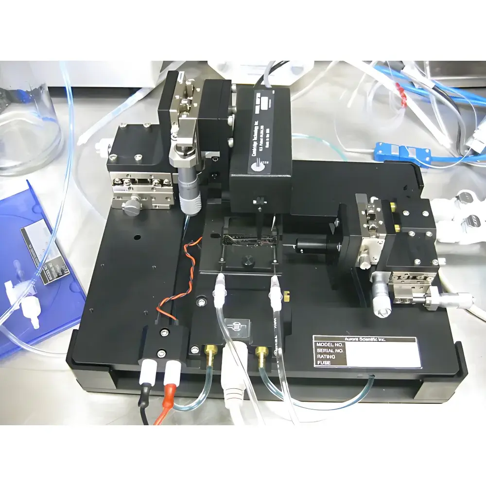
The Aurora 1510A Cardiac & Smooth Muscle Mechanical Testing System is a precision-engineered platform designed for quantitative biomechanical characterization of intact, millimeter-scale striated and smooth muscle preparations—including papillary muscles, ventricular trabeculae, arterial rings, ileal strips, and engineered cardiac microtissues. Based on the principle of servo-controlled length–force feedback, the system enables real-time isometric, isotonic, and work-loop protocols under physiologically relevant conditions (temperature-, pH-, and oxygen-controlled bath environment). Its modular architecture integrates high-fidelity force transduction (sub-micron resolution), rapid-length actuation (<1 ms step response), and synchronized data acquisition to support fundamental research in excitation–contraction coupling, contractile kinetics, passive viscoelasticity, and load-dependent energetics—key parameters required for translational studies in heart failure, vascular dysfunction, and pharmacological screening.
Key Features
- Modular three-configuration platform: Enables flexible adaptation for isolated tissue strips, ring segments, or suspended fiber preparations without hardware re-engineering.
- Integrated XYZ micrometer-stage (1 µm resolution): Precisely aligns the force transducer cantilever, high-speed length controller, and dual-mode sensor relative to the tissue mounting axis—critical for minimizing off-axis loading artifacts.
- Temperature-regulated physiological bath (range: 4–42 °C, ±0.1 °C stability): Equipped with gas-permeable membrane interface and integrated O2/CO2 inlet ports for normoxic or hypoxic superfusion during oxygen consumption assays.
- High-bandwidth force transduction: Piezoresistive sensor with sub-10 µN noise floor and linear response up to 50 mN; calibrated traceably to NIST standards.
- Electromagnetic length controller: Capable of sinusoidal, ramped, or step displacements at frequencies up to 200 Hz with <1% harmonic distortion.
- Unified digital control architecture: All actuators and sensors operate under deterministic real-time scheduling (≤100 µs loop latency) via FPGA-based motion controller.
Sample Compatibility & Compliance
The 1510A accommodates native and bioengineered tissues ranging from 0.5 mm to 8 mm in length and 0.1–2.0 mm in cross-sectional dimension. Mounting is achieved via custom-designed stainless-steel hooks inserted through tissue-specific slits in the bath lid, ensuring reproducible sarcomere alignment and minimal shear stress at attachment points. The system conforms to ISO 13485 design controls for research-use-only instrumentation and supports GLP-compliant audit trails when paired with optional FDA 21 CFR Part 11–enabled software modules. Bath geometry and optical access are optimized for compatibility with upright and inverted research-grade microscopes (including DIC, phase contrast, and confocal systems), enabling simultaneous structural–functional correlation.
Software & Data Management
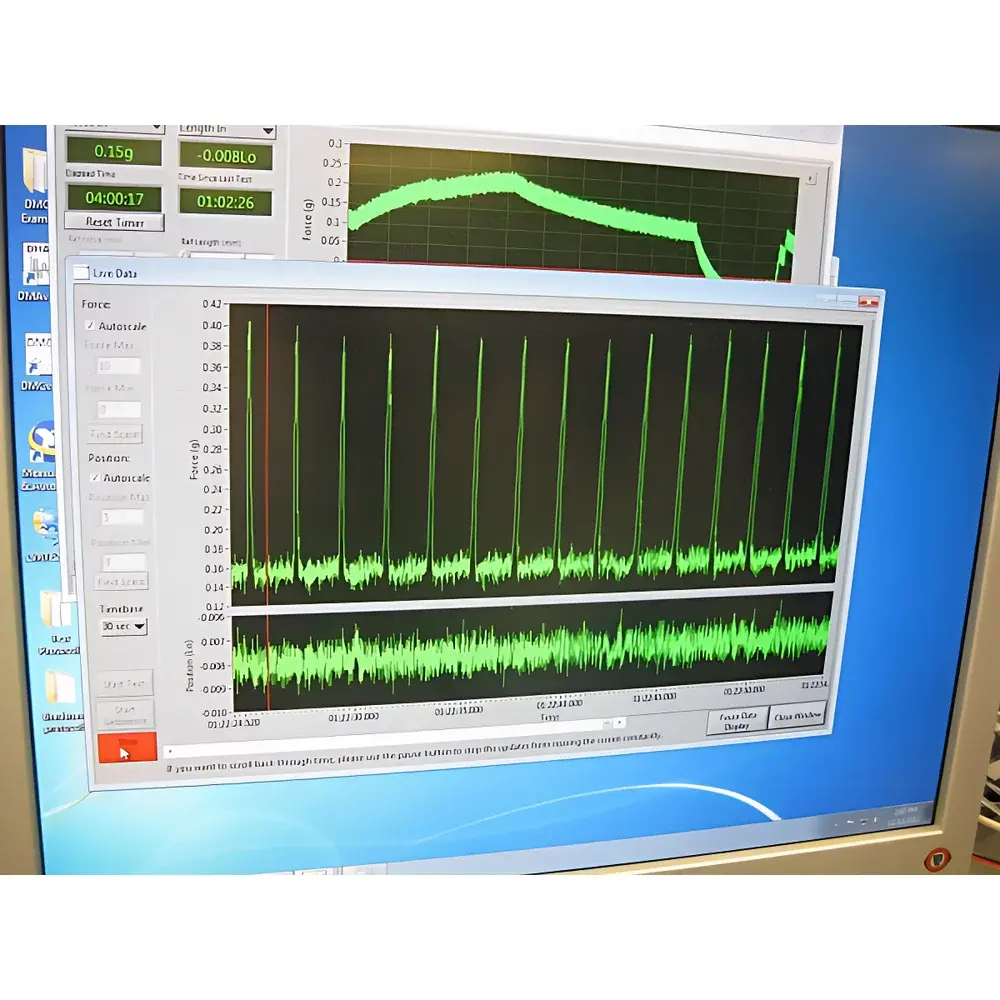
Control and analysis are performed using Aurora’s proprietary MuscleLab v5.x suite—a MATLAB-based application with preconfigured protocols for length–tension, force–velocity, power output, and dynamic stiffness measurements. Raw analog signals (force, length, stimulus timing, bath temperature) are acquired at 10 kHz with 16-bit resolution and timestamped to microsecond precision. The software supports automated curve fitting (Hill equation, exponential relaxation models), export to HDF5/CSV for third-party statistical packages (R, Python SciPy), and direct integration with fluorescence imaging platforms via TTL-synchronized trigger I/O. Optional SarcomereTrack Pro module enables real-time sarcomere length quantification from high-speed video feeds, while calcium imaging add-ons support ratiometric (Fura-2) or single-wavelength (Fluo-4) signal synchronization with mechanical output.
Applications
- Characterization of contractile deficits in genetically modified murine models of hypertrophic cardiomyopathy.
- Assessment of vascular smooth muscle tone modulation by GPCR-targeted therapeutics.
- Quantification of cross-bridge cycling kinetics under varying [Ca2+]i and ATP concentrations.
- Evaluation of biomaterial–myocyte interface mechanics in cardiac patch engineering.
- Validation of computational models of myocardial tissue behavior (e.g., active strain energy density, viscoelastic relaxation time constants).
FAQ
Is the 1510A compatible with oxygen consumption (respirometry) measurements?
Yes—the bath design includes dedicated ports for Clark-type oxygen electrodes and integrates seamlessly with Oroboros O2k or Seahorse XF respirometers via shared temperature and perfusion control interfaces.
Can the system be used for human atrial tissue biopsies?
Yes, provided specimens meet minimum size requirements (≥0.8 mm length, ≥0.15 mm width); standard mounting protocols have been validated in IRB-approved ex vivo studies.
Does the system support closed-loop feedback protocols such as constant-load afterload or simulated arterial impedance?
Yes—real-time PID control loops can be programmed within MuscleLab to emulate physiological loading conditions, including pressure–volume relationships and Windkessel-type afterload waveforms.
What calibration documentation is supplied?
Each unit ships with NIST-traceable force and displacement calibration certificates, ISO/IEC 17025-accredited verification reports, and full uncertainty budgets per parameter.
Is remote operation supported?
Yes—secure SSH-enabled remote desktop access and REST API endpoints allow for protocol scripting, data retrieval, and diagnostic monitoring from external networks (with institutional firewall configuration).


