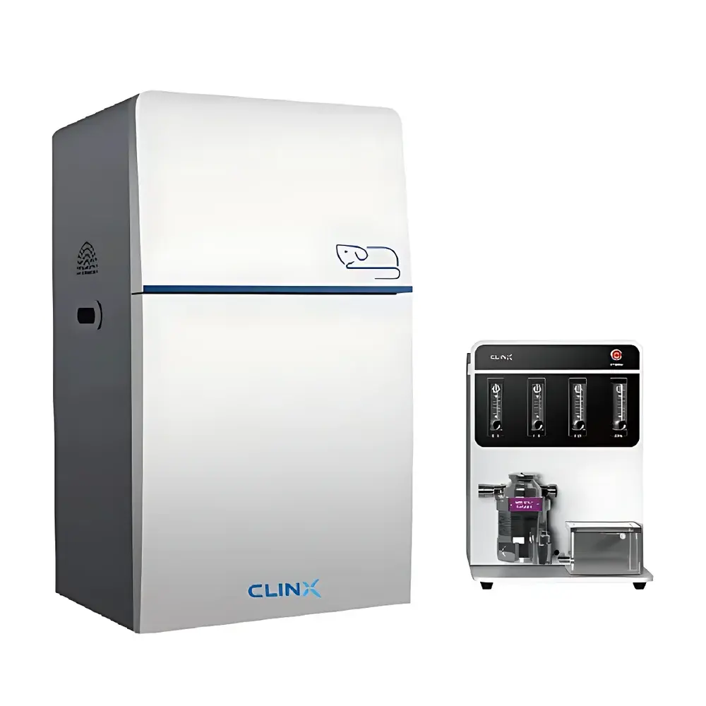
Clinox IVScope 8500 Small Animal In Vivo Optical Imaging System
| Brand | Clinox |
|---|---|
| Origin | Shanghai, China |
| Model | IVScope 8500 |
| Imaging Modality | Bioluminescence & Fluorescence Optical Imaging |
| Camera | 1 MP Front-Illuminated Cooled CCD (Optional Water-Cooled) |
| Cooling Temperature | Down to –90 °C (with optional chiller) |
| Lens | Motorized f/0.8 Wide-Aperture Lens |
| Field of View | Adjustable from 11 cm × 11 cm to 25 cm × 25 cm |
| Maximum FOV | 24 cm × 24 cm |
| Spatial Resolution | 1024 × 1024 pixels |
| Minimum Exposure Time | <10 ms |
| Sample Capacity | Up to 5 Mice Simultaneously |
| Stage Temperature Control | Ambient to 42 °C |
| Excitation Sources | White Light (Reflectance), Optional UV/Vis/NIR LED Arrays & Upconversion Laser Module |
| Filter Wheel | Motorized, Supports Multiple Emission Filters |
| Compliance | Designed for GLP-compliant preclinical research environments |
Overview
The Clinox IVScope 8500 is a high-sensitivity, multi-modal small animal in vivo optical imaging system engineered for quantitative bioluminescence and fluorescence detection in live rodents. Based on cooled charge-coupled device (CCD) photon detection technology, the system captures low-intensity light emissions—such as those from luciferase-expressing tumor cells or NIR-fluorescent probes—within intact biological tissue under physiological conditions. Its optical architecture integrates a deep-cooled front-illuminated CCD sensor, an f/0.8 motorized lens with auto-focusing capability, and a light-tight imaging chamber with precision thermal regulation. Unlike confocal or multiphoton microscopes, the IVScope 8500 operates on wide-field planar imaging principles, enabling rapid, non-invasive longitudinal monitoring of dynamic biological processes—including tumor growth kinetics, immune cell trafficking, pathogen dissemination, and therapeutic biodistribution—across cohorts of up to five mice per acquisition.
Key Features
- Deep-cooled CCD camera (–90 °C operational temperature with optional water-chilled recirculator) ensures ultra-low dark current and high signal-to-noise ratio for sub-picomolar-level bioluminescent detection.
- f/0.8 motorized wide-aperture lens maximizes photon collection efficiency across full spectral range (300–900 nm), critical for weak fluorescence signals and narrow-band emission filters.
- Adjustable field-of-view (11 × 11 cm to 25 × 25 cm) supports both high-resolution localized imaging and whole-body survey modes without mechanical repositioning.
- Integrated anesthesia-compatible stage with precise temperature control (ambient to 42 °C) maintains homeothermic stability during prolonged acquisitions, minimizing physiological stress artifacts.
- Modular excitation source platform includes standard white-light reflectance illumination plus configurable LED arrays (UV, visible, NIR) and an upconversion laser module for rare-earth-doped nanoparticle activation.
- Motorized 6-position filter wheel enables automated spectral unmixing and sequential multi-channel acquisition—essential for multiplexed reporter studies and autofluorescence subtraction protocols.
Sample Compatibility & Compliance
The IVScope 8500 accommodates standard laboratory rodent models including nude, NSG, C57BL/6, BALB/c, and transgenic strains weighing ≤35 g. Its ergonomic stage design permits reproducible positioning of mice in supine or prone orientation, with optional restraint accessories for head-fixed or lateral-view imaging. The system conforms to ISO 13485-aligned manufacturing practices and is validated for use in GLP-regulated preclinical pharmacology and toxicology studies. All hardware and software components support audit-trail generation per FDA 21 CFR Part 11 requirements when deployed with Clinox’s optional secure user authentication and electronic signature modules. Data integrity is maintained via timestamped raw image storage (TIFF/FITS format), metadata embedding (exposure time, binning, filter ID, temperature), and lossless compression protocols compliant with MIAME and MINSEQE reporting standards.
Software & Data Management
Acquisition and analysis are performed using Clinox ImageStudio Pro v5.x—a dedicated, scriptable platform supporting batch processing, region-of-interest (ROI) quantification, kinetic curve fitting, and spectral deconvolution. The software implements standardized calibration routines using NIST-traceable luminance references and supports export to HDF5, MATLAB (.mat), and CSV formats for downstream statistical modeling in R or Python. Integrated tools include background subtraction algorithms optimized for tissue autofluorescence, automatic animal contour segmentation using adaptive thresholding, and longitudinal registration for cross-session anatomical alignment. Audit logs record all user actions—including parameter modifications, ROI edits, and export events—with immutable timestamps and operator IDs. Raw data files are stored with embedded DICOM-SR headers for PACS interoperability in translational research centers.
Applications
- Oncology: Quantitative tracking of orthotopic tumor burden, metastatic spread, and response to checkpoint inhibitors or ADC therapies using dual-luciferase reporters.
- Immunology: Real-time visualization of adoptively transferred T cells or macrophages labeled with near-infrared fluorophores in models of colitis, EAE, or graft-versus-host disease.
- Infectious Disease: Spatiotemporal mapping of bacterial load (e.g., Salmonella, Staphylococcus) or viral replication (e.g., influenza, SARS-CoV-2 pseudoviruses) via lux/fluorophore-tagged pathogens.
- Neuroscience: Monitoring neuroinflammation via TSPO-targeted tracers or neuronal activity using genetically encoded calcium indicators (e.g., GCaMP).
- Regenerative Medicine: Engraftment and migration kinetics of mesenchymal stem cells or iPSC-derived progenitors labeled with membrane-intercalating dyes or luciferase fusion constructs.
- Drug Development: PK/PD correlation of fluorescently conjugated therapeutics, nanoparticle biodistribution, and target engagement validation in disease-relevant models.
FAQ
What is the minimum detectable photon flux for bioluminescence imaging?
The system achieves a theoretical detection limit of ~103 photons/sec/cm2/sr under optimal cooling (–90 °C) and longest exposure settings; actual sensitivity depends on emission wavelength, tissue depth, and background autofluorescence.
Can the IVScope 8500 perform spectral unmixing for overlapping fluorophores?
Yes—using the motorized filter wheel and multi-emission acquisition mode, users can collect ≥3 spectral channels and apply linear unmixing algorithms within ImageStudio Pro to resolve fluorophores with <100 nm separation (e.g., Cy5.5/Cy7).
Is anesthesia integration supported?
The system includes ports and mounting interfaces for third-party isoflurane vaporizers and nose-cone delivery systems; temperature-controlled stage operation remains stable during continuous gas anesthesia.
Does the software support compliance with regulatory submissions?
When configured with 21 CFR Part 11 add-ons, ImageStudio Pro provides electronic signatures, role-based access control, and immutable audit trails required for IND-enabling toxicology reports and CMC documentation.
What maintenance is required for long-term performance stability?
Annual recalibration of camera quantum efficiency and lens transmission profiles is recommended; Clinox offers certified service contracts including dark-frame characterization, filter spectral verification, and vacuum seal integrity testing.


