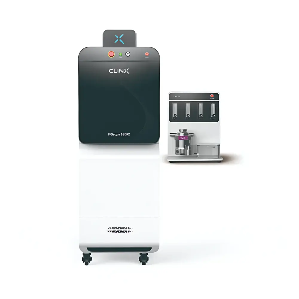
Clinox IVScope 8500X Multi-Modal X-ray and Optical Small Animal In Vivo Imaging System
| Brand | Clinox |
|---|---|
| Origin | Shanghai, China |
| Model | IVScope 8500X |
| Imaging Modality | Optical (Bioluminescence/Fluorescence) + X-ray |
| Max Sample Capacity | 5 mice |
| CCD Resolution | 1024 × 1024 |
| Field of View | 11 cm × 11 cm to 25 cm × 25 cm (continuously adjustable) |
| X-ray Minimum Resolvable Feature Size | 0.16 mm |
| Lens Aperture | f/0.8 motorized auto-focus lens |
| Radiation Shielding | ≤0.5 mR/h at 5 cm from chassis wall (compliant with FDA 21 CFR 1020.40) |
| Stage Temperature Control | Ambient to 42 °C |
| Camera Type | Front-illuminated CCD (water-cooling optional) |
| Excitation Sources | White light reflectance (standard), UV/VIS/NIR reflectance & upconversion laser modules (optional) |
| Filter System | Motorized filter wheel with emission filter support |
| Anesthesia Interface | Integrated gas anesthesia delivery |
Overview
The Clinox IVScope 8500X is a preclinical multi-modal in vivo imaging platform engineered for simultaneous high-fidelity optical (bioluminescence and fluorescence) and X-ray radiographic imaging in small animal models. It operates on the principle of complementary modality fusion: bioluminescent and fluorescent signals report molecular and cellular activity—such as gene expression, protease activity, or cell trafficking—while X-ray imaging provides anatomical context through differential X-ray attenuation by bone, soft tissue, and contrast agents. This dual-data acquisition enables spatial co-registration of functional and structural information without requiring sequential repositioning or image registration post-acquisition. The system employs a vacuum-cooled front-illuminated CCD detector optimized for low-light detection, coupled with an f/0.8 motorized lens assembly to maximize photon collection efficiency under ultra-low signal conditions. X-ray imaging is performed using a pulsed, low-dose microfocus source with real-time dose monitoring and hardware-based beam collimation, ensuring compliance with international radiation safety standards for laboratory environments.
Key Features
- Multi-modal data acquisition: Synchronized bioluminescence, fluorescence, and X-ray imaging in a single session
- High-sensitivity cooled CCD detector: Configurable for −90 °C operation via optional water-circulation chiller to suppress dark current and enhance signal-to-noise ratio
- f/0.8 motorized lens with auto-focus and variable field-of-view (11 × 11 cm to 25 × 25 cm), enabling rapid adaptation across anatomical scales—from whole-body murine imaging to localized organ-level resolution
- Integrated gas anesthesia manifold compatible with isoflurane delivery and real-time physiological monitoring ports (optional)
- Thermally regulated specimen stage (ambient to 42 °C) supporting longitudinal studies requiring controlled body temperature maintenance
- Modular excitation architecture: Standard white-light reflectance; optional UV (365 nm), visible (470–630 nm), NIR (740–850 nm), and upconversion laser (980 nm) sources for advanced probe activation
- Motorized emission filter wheel accommodating up to 6 position-specific bandpass filters for spectral unmixing and multiplexed probe detection
Sample Compatibility & Compliance
The IVScope 8500X accommodates live rodents (mice and rats) weighing up to 50 g, with support for up to five animals per imaging session via programmable stage positioning and independent ROI selection. All X-ray components meet FDA 21 CFR 1020.40 requirements for cabinet X-ray systems: measured exposure rate does not exceed 0.5 mR/h at 5 cm from any external surface. The system is designed for use in GLP-compliant laboratories and supports audit-ready documentation workflows. While not certified to ISO 13485, its mechanical and electrical architecture adheres to IEC 61010-1 safety standards for laboratory equipment. Optional integration with third-party physiological monitors (e.g., ECG, respiration, temperature) allows correlation of imaging endpoints with functional parameters during acquisition.
Software & Data Management
Acquisition and analysis are managed through Clinox ImageStudio Pro—a validated software suite supporting DICOM export, TIFF/PNG sequence generation, and quantitative ROI-based photonic flux measurement (photons/sec/cm²/sr). The software implements background subtraction algorithms based on rolling-ball and morphological filtering, and includes spectral unmixing tools for fluorophore separation when multiple probes are used. All user actions—including exposure time, binning mode, filter selection, and region annotation—are logged with timestamps and operator ID in an embedded audit trail compliant with ALCOA+ principles. Raw data files are stored in vendor-neutral HDF5 format, enabling interoperability with MATLAB, Python (via h5py), and open-source analysis pipelines such as QuantiFly or FIJI/ImageJ plugins. Export options include CSV tables for statistical analysis and annotated PDF reports suitable for regulatory submissions.
Applications
- Oncology: Longitudinal tracking of tumor burden, metastasis, and therapeutic response using luciferase-expressing xenografts combined with skeletal X-ray assessment of osteolytic lesions
- Inflammation & immunology: Quantification of immune cell infiltration via near-infrared fluorescent probes co-registered with joint or lung anatomy
- Gene therapy validation: Spatial mapping of transgene expression relative to anatomical landmarks in liver, muscle, or CNS
- Drug biodistribution: Co-localization of fluorescently labeled therapeutics with soft-tissue contrast or bone density changes
- Toxicology: Assessment of hepatomegaly or pulmonary edema via X-ray densitometry paired with apoptosis-associated luminescent reporters
<liCardiovascular research: Monitoring of cardiac reporter activity alongside X-ray visualization of calcification or stent placement
FAQ
Is the IVScope 8500X compliant with FDA 21 CFR Part 11 for electronic records and signatures?
No—Part 11 applies primarily to clinical trial systems and manufacturing quality records. However, ImageStudio Pro supports audit-trail logging, user authentication, and electronic signature-capable reporting modules that may be configured to align with internal SOPs for regulated preclinical studies.
Can the system perform kinetic bioluminescence imaging over extended durations?
Yes—software-defined acquisition sequences support time-lapse protocols up to 72 hours, with automated stage repositioning and adaptive exposure control to maintain dynamic range across varying signal intensities.
What is the maximum permissible X-ray tube voltage and current for this system?
The integrated X-ray source operates at a fixed kVp of 40 kV and maximum tube current of 0.5 mA, optimized for soft-tissue contrast in murine models while minimizing dose accumulation.
Does the system support intravital fluorescence imaging with confocal or two-photon capability?
No—IVScope 8500X is a widefield epifluorescence and transmission X-ray platform. Confocal or multiphoton imaging requires dedicated upright or inverted microscope systems with scanning optics.
Is remote operation supported for biosafety level 2 (BSL-2) containment environments?
Yes—full software control, including acquisition triggering and parameter adjustment, can be executed via Ethernet-connected client workstations located outside the imaging suite, minimizing operator exposure during infectious agent studies.


