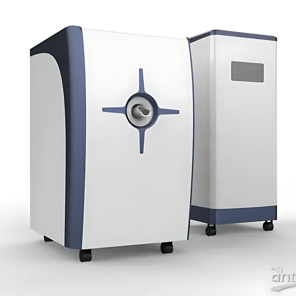
HTNMR HT-MRSI40-60KY 1.0T Preclinical MRI System for Small Animal Imaging
| Brand | HTNMR |
|---|---|
| Origin | Shanghai, China |
| Manufacturer Type | Direct Manufacturer |
| Instrument Type | Preclinical Magnetic Resonance Imaging (MRI) System |
| Model | HT-MRSI40-60KY |
| Magnetic Field Strength | 1.0 T ± 0.05 T |
| Proton Larmor Frequency | 42 MHz ± 2 MHz |
| Magnet Bore Diameter | 300 mm |
| Effective Sample Volume (RF Coil) | Φ60 mm × H80 mm |
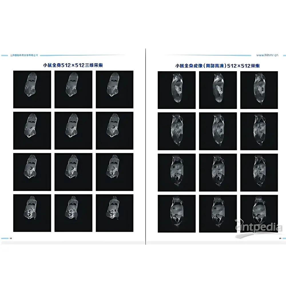
| Spatial Resolution (Max) | 0.05 mm (measured with 0.05 mm water phantom) |
| Gradient Strength | 100 mT/m (X/Y/Z) |
| Field Homogeneity | < 8 ppm over 50 mm × 50 mm × 80 mm DSV |
| Image Linearity | > 98% in X/Y/Z (50 mm × 50 mm × 80 mm) |
| Temporal Stability | < 100 Hz/h Larmor frequency drift |
| Temperature Control Stability | ±0.005 °C (cavity), display resolution 1 m°C |
| Image Matrix | Up to 256 × 256 × 128 |
| DICOM Compliance | Yes |
| Sensitivity | 52 dB |
| Resolution | 98% |
Overview
The HTNMR HT-MRSI40-60KY is a 1.0 tesla preclinical magnetic resonance imaging system engineered for high-fidelity structural, functional, and metabolic studies in small animal models—including rats, mice, and other rodent-sized specimens. Based on superconducting magnet technology and pulsed RF excitation principles, the system operates at a proton Larmor frequency of 42 MHz and delivers quantitative MR signal acquisition through spin-echo, gradient-echo, and inversion-recovery pulse sequences. Its open-bore 300 mm magnet architecture accommodates dedicated RF coils optimized for Φ60 mm × H80 mm sample volumes, enabling whole-body imaging of adult rats while maintaining sub-100 µm spatial resolution under optimized acquisition conditions. Designed for longitudinal in vivo studies, the system supports dynamic contrast-enhanced MRI, T1/T2 relaxometry, diffusion-weighted imaging, and spectroscopic imaging protocols—making it suitable for translational research in oncology, neurodegeneration, cardiovascular disease, and pharmacokinetic evaluation.
Key Features
- Stable 1.0 T permanent or resistive magnet (±0.05 T field tolerance) with homogeneity < 8 ppm over a 50 mm × 50 mm × 80 mm DSV (Diameter Spherical Volume)
- Dedicated quadrature or volume RF coil configuration supporting Φ60 mm × H80 mm specimen dimensions, compatible with physiological monitoring accessories (respiratory gating, ECG leads, temperature probes)
- Triple-axis gradient system with maximum strength of 100 mT/m and linearity > 98% across X/Y/Z directions within the imaging volume
- High-precision cavity temperature control (±0.005 °C stability, 1 m°C display resolution) for thermally sensitive experiments including diffusion and fMRI
- Real-time field-frequency lock and active shimming to maintain temporal stability (< 100 Hz/h Larmor drift), critical for multi-session longitudinal studies
- Full pulse sequence library compliant with standard NMR/MRI conventions: Spin Echo (SE), Turbo Spin Echo (TSE), Gradient Echo (GRE), Inversion Recovery (IR), CPMG, and 3D volumetric acquisitions
Sample Compatibility & Compliance
The HT-MRSI40-60KY accommodates live rodents (e.g., Sprague-Dawley rats up to 500 g, C57BL/6 mice), excised tissues, tissue phantoms, and biomaterials. It supports non-invasive in vivo imaging under anesthesia (isoflurane/O2 delivery integration optional), as well as ex vivo perfusion-fixed specimens. All image data are stored in DICOM 3.0 format, ensuring interoperability with PACS systems, third-party analysis platforms (e.g., Horos, 3D Slicer), and regulatory-compliant archival workflows. The system meets essential electromagnetic compatibility (EMC) requirements per IEC 61326-1 and adheres to safety guidelines outlined in ISO/IEC 17025 for analytical instrument validation. While not FDA-cleared for clinical diagnostics, its architecture aligns with GLP/GMP documentation practices for preclinical imaging studies intended for IND-enabling submissions.
Software & Data Management
The proprietary acquisition and reconstruction software provides graphical pulse sequence editing, real-time k-space monitoring, and automated parameter optimization for SNR and contrast tuning. Raw k-space data are preserved alongside reconstructed NIfTI and DICOM files, enabling offline processing with MATLAB, Python (NiBabel, nibabel), or FSL. Audit trails record operator identity, acquisition timestamps, sequence parameters, and hardware calibration logs—supporting traceability requirements under 21 CFR Part 11 when deployed in regulated environments. Batch processing tools facilitate multi-subject analysis, ROI-based quantification, and T1/T2 map generation. Export modules support CSV, MATLAB .mat, and JSON formats for integration into statistical pipelines (e.g., R, SPSS, GraphPad Prism).
Applications
- Longitudinal tumor growth monitoring and anti-angiogenic therapy assessment in murine xenograft models
- Structural neuroimaging of transgenic mouse models of Alzheimer’s disease, including hippocampal volumetry and white matter integrity mapping
- Cardiac function evaluation via cine-MRI and late gadolinium enhancement (LGE) in rat myocardial infarction models
- In vivo relaxometry for nanoparticle-based contrast agent development (r1/r2 quantification)
- Molecular imaging using hyperpolarized 13C substrates (with optional cryoprobe and RF upgrade)
- Material science applications including polymer hydration dynamics, hydrogel swelling behavior, and porous media characterization
FAQ
Is this system suitable for functional MRI (fMRI) in rodents?
Yes—the system supports echo-planar imaging (EPI) and gradient-echo sequences with temporal resolution sufficient for BOLD fMRI in anesthetized rats when combined with physiological monitoring.
Can the system be upgraded for spectroscopy (MRS)?
Yes—optional broadband RF amplifiers, dual-tuned coils (e.g., 1H/31P), and spectral acquisition modules enable localized single-voxel and chemical shift imaging (CSI) protocols.
What level of technical support and service coverage is provided?
HTNMR offers on-site installation, operator training, annual preventive maintenance, and remote diagnostics. Extended warranty and service contracts include magnet shimming recalibration and gradient performance verification.
Does the system comply with international data standards for publication and regulatory submission?
All acquired images conform to DICOM 3.0 Part 10 file structure, including embedded metadata (acquisition parameters, scanner model, field strength). Raw k-space data export enables reproducible reconstruction and third-party validation per MIAME/MINIMUM guidelines.
What are the facility requirements for installation?
A dedicated shielded room (RF and magnetic shielding), stable 220 V ± 5%, 50 Hz power supply with dedicated grounding, ambient temperature control (20–25 °C), and vibration-isolated floor mounting are required. Site survey and shielding design assistance are included in the commissioning package.