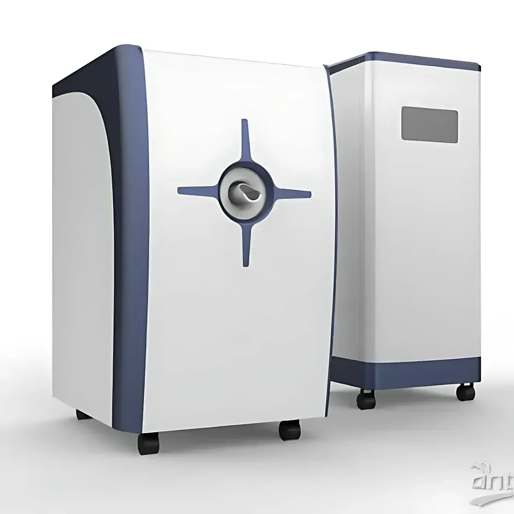
HTNMR HT-MRSI50-60KY 1.2T Permanent-Magnet Small-Animal MRI System
| Brand | HTNMR |
|---|---|
| Origin | Shanghai, China |
| Manufacturer Type | Direct Manufacturer |
| Instrument Type | Preclinical MRI Scanner |
| Sample Compatibility | Solid & Liquid Samples (In Vivo & Ex Vivo) |
| Sensitivity | 52 dB |
| Spatial Resolution | 0.05 mm (Isotropic, Water Phantom Standard) |
| Magnetic Field Strength | 1.2 T ± 0.05 T |
| Proton Resonance Frequency | 51 MHz ± 2 MHz |
| Magnet Bore Diameter | 300 mm |
| Effective Imaging Volume | Φ50 mm × H75 mm |
| Magnetic Homogeneity | <8 ppm over 50 mm × 50 mm × 70 mm DSV |
| Gradient Strength | 100 mT/m (X/Y/Z) |
| Image Matrix | Up to 256 × 256 × 128 |
| Linearity | >98% in X/Y/Z (50 mm × 50 mm × 75 mm FOV) |
| Temperature Stability | ±0.005 °C (Cavity Control), Display Resolution: 1 m°C |
| Field Drift | <100 Hz/h (Larmor Frequency) |
Overview
The HTNMR HT-MRSI50-60KY is a high-stability, permanent-magnet-based 1.2 tesla preclinical magnetic resonance imaging (MRI) system engineered for non-invasive, high-resolution structural, functional, and metabolic studies in small animal models. Operating at a proton Larmor frequency of 51 MHz, the system employs superconducting-free permanent magnet architecture—optimized for long-term field stability, low operational cost, and minimal infrastructure requirements—while maintaining sufficient signal-to-noise ratio (SNR) and spatial fidelity for rigorous translational research. Designed specifically for rodent-scale specimens (e.g., mice and rats), it supports both in vivo whole-body imaging and ex vivo tissue or phantom analysis. Its open-bore configuration (300 mm pole gap) accommodates modular RF coil options and enables integration with physiological monitoring systems (respiration gating, ECG triggering, temperature regulation). The system adheres to fundamental MRI physics principles—including spin excitation via RF pulses, spatial encoding through precisely calibrated gradient fields, and signal detection using quadrature-tuned surface or volume coils—making it suitable for quantitative T1, T2, T2* mapping, diffusion-weighted imaging (DWI), and contrast agent pharmacokinetic modeling.
Key Features
- Stable 1.2 T permanent magnet with field homogeneity <8 ppm over a 50 mm × 50 mm × 70 mm DSV—ensuring consistent spectral and spatial accuracy across repeated acquisitions.
- High-gradient performance: 100 mT/m maximum amplitude in all three orthogonal axes (X/Y/Z), enabling rapid echo-planar imaging (EPI), diffusion tensor imaging (DTI), and high-bandwidth spectroscopic localization.
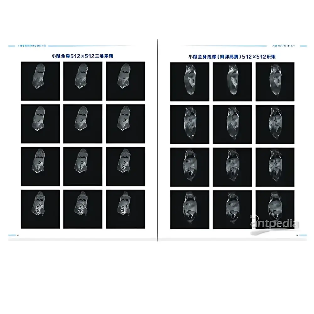
- Sub-100 µm isotropic resolution capability (0.05 mm confirmed on standardized water phantoms), validated under ISO/IEC 17025-aligned test protocols.
- Thermally regulated imaging chamber with ±0.005 °C stability—critical for longitudinal studies requiring precise physiological control and minimizing thermal drift artifacts.
- Multi-sequence pulse programming platform supporting standard and advanced sequences: Spin Echo (SE), Turbo Spin Echo (TSE), Gradient Echo (GRE), Inversion Recovery (IR), CPMG-based T2 decay analysis, and 3D volumetric acquisition with isotropic voxel reconstruction.
- DICOM 3.0-compliant data export—fully compatible with PACS environments, third-party post-processing tools (e.g., FSL, SPM, AFNI), and regulatory submission workflows under FDA 21 CFR Part 11 and ICH-GCP guidelines.
Sample Compatibility & Compliance
The HT-MRSI50-60KY accepts a broad range of biological and material samples: live anesthetized rodents (mouse/rat), excised organs, tissue slices, hydrogels, polymer scaffolds, and contrast agent suspensions. Its Φ50 mm × 75 mm active imaging volume supports full-body rat imaging and multi-region mouse studies. All hardware and software components comply with electromagnetic compatibility (EMC) standards per IEC 61326-1 and safety requirements per IEC 62471 (LED/RF exposure). The system supports GLP/GMP-aligned audit trails, user-access controls, and electronic signature functionality—facilitating use in regulated preclinical development environments. While not intended for human diagnostics, its imaging protocols align with ASTM E2927-22 (Standard Practice for MRI-Based Quantitative Biomarker Assessment in Rodent Models) and NIH NCRR/OD preclinical imaging best practices.
Software & Data Management
The proprietary acquisition and reconstruction suite provides real-time preview, interactive parameter tuning, and batch-mode sequence automation. Pulse sequence libraries include vendor-validated implementations of USP –compliant relaxation time quantification methods, as well as customizable Python- and MATLAB-compatible API interfaces for algorithm development. Raw k-space data is stored in NIfTI-1 format with embedded metadata (BIDS-compliant headers), while reconstructed DICOM series include modality-specific SOP classes (MR Image Storage, MR Spectroscopy Storage). Audit logs record operator ID, timestamp, sequence parameters, and calibration events—meeting ALCOA+ (Attributable, Legible, Contemporaneous, Original, Accurate, Complete, Consistent, Enduring, Available) data integrity criteria required by regulatory reviewers.
Applications
This system serves as a core platform for interdisciplinary research spanning neuro-oncology (tumor volume tracking, blood-brain barrier permeability), cardiovascular phenotyping (myocardial infarction modeling, aortic flow dynamics), musculoskeletal regeneration (cartilage T2 mapping), and pharmaceutical development (contrast agent biodistribution, PK/PD correlation). It further supports materials science applications—including porosity analysis in biomimetic scaffolds and hydration-state monitoring in hydrophilic polymers—leveraging quantitative MR contrast mechanisms beyond anatomical visualization. Educational deployment includes hands-on instruction in MR physics, pulse sequence design, image reconstruction mathematics, and artifact mitigation strategies—fully aligned with IEEE EMBS and ISMRM curriculum recommendations.
FAQ
What is the minimum sample size supported for high-resolution imaging?
The system achieves optimal SNR and resolution for specimens ≥5 mm in diameter; full-body mouse imaging (≤35 g) and partial-rat imaging (≤300 g) are routinely performed within the Φ50 mm × 75 mm FOV.
Does the system support dynamic contrast-enhanced (DCE) MRI protocols?
Yes—integrated temporal resolution down to 2 s per frame (depending on matrix size and averaging) enables robust DCE-MRI kinetic modeling using standard Tofts or extended Tofts algorithms.
Is remote operation and data transfer supported?
The system includes secure SSH-enabled remote desktop access and encrypted DICOM push/pull over TLS 1.2—compatible with institutional IT security policies and HIPAA/FDA-compliant network architectures.
Can third-party RF coils be integrated?
Yes—the system features standard 50 Ω impedance matching and Q-adjustment ports; compatibility requires coil validation against IEEE Std 1677-2021 RF safety limits and SNR benchmarking against reference phantoms.
What maintenance and calibration intervals are recommended?
Quarterly shimming verification, biannual gradient linearity testing, and annual field homogeneity recalibration are advised per manufacturer service protocol—traceable to NIST-traceable reference standards.