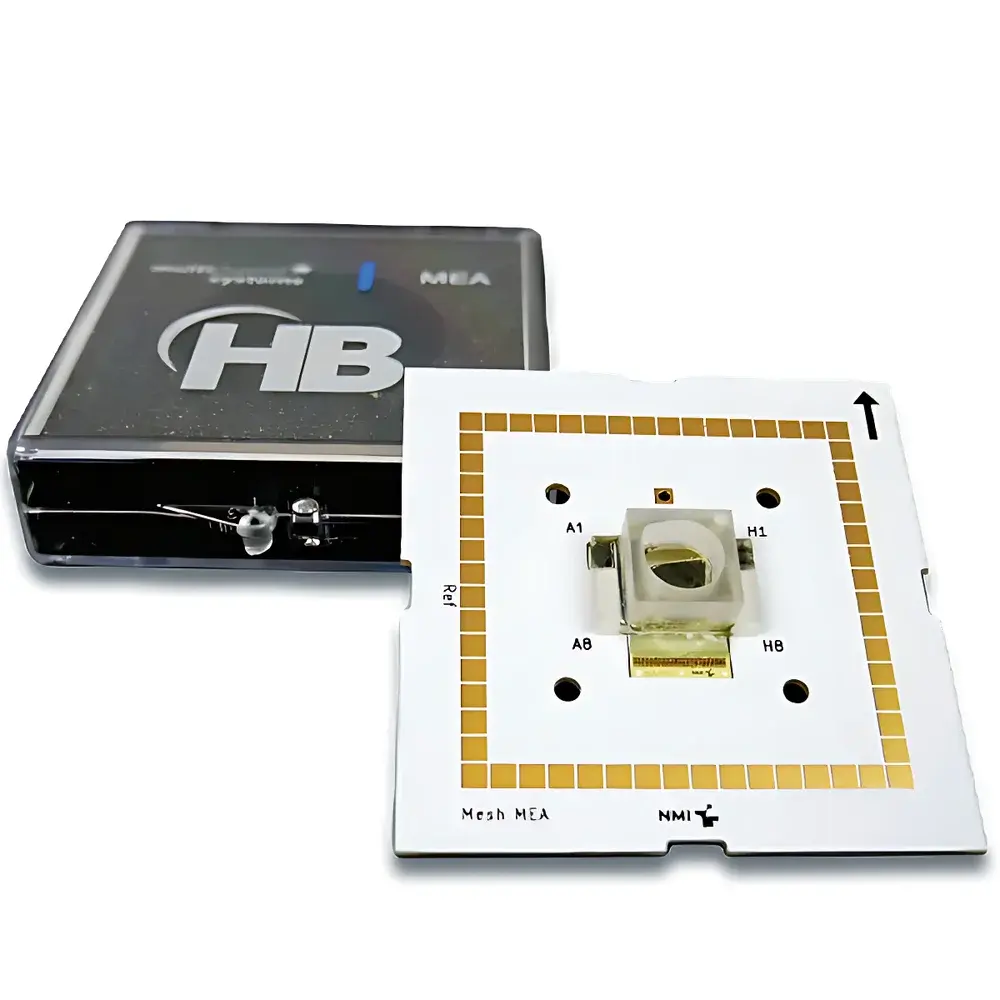
Multi Channel Systems Mesh MEA – 60-Channel Flexible 3D Microelectrode Array for Organoid Electrophysiology
| Brand | Multi Channel Systems |
|---|---|
| Origin | Germany |
| Manufacturer | Yes |
| Import Status | Imported |
| Model | Mesh MEA |
| Instrument Type | Single-well |
| Application | In Vitro |
| Electrode Spacing | 200 µm |
| Electrode Diameter | 30 µm |
| Electrode Layout | Mesh-type MEA |
| Number of Electrodes | 60 |
| Dimensions | 49 mm × 49 mm |
Overview
The Multi Channel Systems Mesh MEA is a purpose-engineered 3D microelectrode array platform designed to overcome fundamental limitations of conventional planar MEAs in organoid electrophysiology. Unlike traditional 2D substrates that restrict signal acquisition to the basal surface, the Mesh MEA integrates 60 titanium nitride (TiN) microelectrodes—each 30 µm in diameter and spaced at 200 µm—into an ultra-thin (7 µm), biocompatible polyimide mesh scaffold. This architecture enables true volumetric integration: human or rodent-derived organoids grow *around* and *through* the mesh, allowing electrodes to reside within the 3D tissue volume. The device operates on the principle of extracellular field potential recording under controlled perfusion, delivering high-fidelity, spatially resolved electrophysiological data from intact, unperturbed organoid structures—without mechanical sectioning, embedding, or electrode insertion.
Key Features
- 60-channel flexible MEA chip embedded in a 7 µm-thick polyimide mesh—engineered for mechanical compliance with dynamic 3D tissue growth
- TiN-coated electrodes (30 µm diameter, 200 µm pitch) optimized for low impedance (<100 kΩ @ 1 kHz) and high signal-to-noise ratio
- Simultaneous sampling at 50 kHz per channel with 24-bit resolution, supporting detection of fast neuronal spikes and slow network oscillations
- Integrated stimulation capability: programmable biphasic voltage or current pulses (±5 V, ±10 mA) for closed-loop interrogation
- Optical transparency across visible and near-UV spectra—fully compatible with live-cell fluorescence imaging, calcium indicators (e.g., GCaMP), and optogenetic actuation
- USB 3.0 interface enabling real-time streaming (>1 Gbps aggregate bandwidth) and deterministic latency (<100 µs jitter)
- Modular I/O architecture: 8 digital TTL inputs/outputs, 4 analog inputs (±10 V, 16-bit), and stereo audio output for synchronized multimodal experiments
Sample Compatibility & Compliance
The Mesh MEA supports long-term (≥28 days) functional recording from diverse 3D neural and non-neural organoid models—including cerebral, hippocampal, midbrain, retinal, cardiac spheroids, pancreatic islet clusters, and biofabricated constructs. Its open-mesh geometry permits unimpeded nutrient/waste exchange and enables gas–liquid interface (GLI) culture configurations critical for cortical layering and vascular mimicry. The system complies with ISO 13485:2016 (medical device quality management) and adheres to GLP-relevant documentation standards. Data acquisition firmware supports audit trails, electronic signatures, and time-stamped metadata export—facilitating alignment with FDA 21 CFR Part 11 requirements for regulated preclinical studies.
Software & Data Management
Controlled via MC_Rack v5.x software suite, the Mesh MEA enables fully customizable experimental workflows: user-defined stimulus protocols, real-time spike sorting (using PCA + k-means clustering), LFP bandpass filtering (0.1–300 Hz), and cross-channel coherence analysis. Raw binary data (.mcdata) are stored with embedded calibration parameters and exported in HDF5 or NWB 2.0 format for interoperability with Python (Neo, MNE), MATLAB (Chronux), and Brainstorm. Cloud-synced project archives include full experimental provenance—stimulus history, environmental logs (temperature, pH, O₂), and operator annotations—ensuring reproducibility and traceability.
Applications
- Neurodevelopmental modeling: longitudinal tracking of network burst dynamics during cortical organoid maturation
- Pharmacological profiling: concentration–response assessment of sodium channel blockers (e.g., lidocaine), NMDA antagonists (e.g., ketamine), or neurotoxicants (e.g., methylmercury)
- Disease-in-a-dish studies: functional phenotyping of patient-derived iPSC organoids in epilepsy, Parkinson’s, or Rett syndrome
- Cardiac safety pharmacology: detection of arrhythmic endpoints (early afterdepolarizations, conduction block) in heart organoids per CiPA guidelines
- Multi-organ interaction: co-culture platforms integrating neural + cardiac or neural + hepatic organoids with inter-chip electrical coupling
FAQ
Can the Mesh MEA be sterilized and reused?
No—the Mesh MEA is a single-use, sterile-packaged device. Autoclaving or ethanol immersion compromises polyimide integrity and TiN electrode stability.
Is the mesh compatible with standard MEA2100 amplifier systems?
Yes—fully backward-compatible with all MEA2100 series amplifiers (MEA2100-2i, MEA2100-60i, MEA2100-60i-USB) and requires no hardware modification.
What perfusion rates are supported without compromising organoid adhesion?
Recommended laminar flow range: 5–20 µL/min using gravity-driven or syringe-pump systems; higher rates may dislodge immature organoids but are tolerated post-day 14.
How is electrode impedance validated prior to recording?
MC_Rack performs automated impedance spectroscopy (1 Hz–100 kHz) at startup; acceptance threshold: <150 kΩ at 1 kHz for all 60 channels.
Does the system support simultaneous calcium imaging and electrophysiology?
Yes—optical transparency and minimal autofluorescence enable concurrent widefield or confocal Ca²⁺ imaging with sub-millisecond temporal registration to electrophysiological traces.
Related Products


