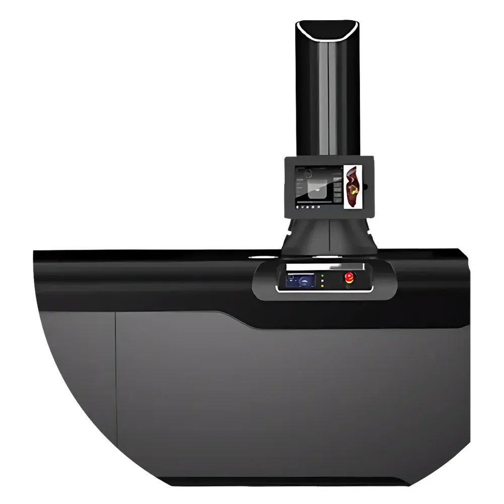
Xerra CFT Cryo-Fluorescence Tomography Imaging System
| Brand | Xerra |
|---|---|
| Origin | USA |
| Manufacturer Type | Authorized Distributor |
| Import Status | Imported |
| Model | CFT |
| Price | USD 1,520,000 (FOB US Port) |
Overview
The Xerra CFT Cryo-Fluorescence Tomography Imaging System is a high-resolution, quantitative optical tomographic platform engineered for ex vivo 3D fluorescence reconstruction of intact small animal specimens, excised tissues, and human biospecimens. Unlike in vivo modalities such as PET, MRI, or CT—which face fundamental limitations in sensitivity, spatial resolution, and molecular specificity—the CFT system operates on the principle of serial cryosectioning coupled with synchronized dual-channel (fluorescence + brightfield) macroscopic imaging. Specimens are embedded in optimal cutting temperature (OCT) compound, frozen to cryogenic temperatures (−20 °C to −30 °C), and sectioned using a motorized cryomicrotome with sub-micron positional feedback. At each slice—down to 20 µm isotropic thickness—the system acquires registered widefield fluorescence images (with excitation/emission filter sets spanning 400–900 nm) and high-fidelity white-light reflectance images. These 2D datasets are then computationally reconstructed into volumetric, isotropic 3D datasets with native voxel resolution of 20 µm × 20 µm × 20 µm, enabling precise anatomical registration, quantitative signal localization, and multi-scale correlation from organ down to cellular compartments.
Key Features
- Isotropic 20 µm volumetric resolution across whole murine specimens (up to 40 mm length) and human tissue blocks (up to 25 mm × 25 mm × 25 mm)
- Automated cryosectioning workflow with real-time Z-stage calibration and thermal drift compensation
- Dual-modality acquisition per slice: fluorescence (7-channel filter wheel, LED-based excitation) + brightfield (LED-illuminated, 12-bit sCMOS sensor)
- Integrated spectral unmixing engine for multi-fluorophore quantification (e.g., GFP/mCherry/iRFP/tdTomato/Cy5.5) with cross-talk correction
- Hardware-synchronized stage control and image capture to ensure <0.5 µm slice-to-slice registration accuracy
- Modular optical path design compliant with ANSI Z80.10 and ISO 10940 standards for fluorescence intensity calibration
Sample Compatibility & Compliance
The CFT system accommodates formalin-fixed paraffin-embedded (FFPE) rehydrated sections, fresh-frozen OCT-embedded tissues, and decalcified bone specimens. It supports standard histological staining (H&E, DAPI, IHC) without signal interference and enables co-registration with downstream confocal or light-sheet microscopy via fiducial marker alignment. All hardware and software components meet CE marking requirements under Directive 2014/30/EU (EMC) and 2014/35/EU (LVD). The acquisition and reconstruction pipeline is validated for GLP-compliant preclinical studies and supports audit trails, electronic signatures, and data integrity features aligned with FDA 21 CFR Part 11 and ISO/IEC 17025:2017 Annex A.2 for analytical instrument qualification.
Software & Data Management
- Xerra CFT Studio v4.2: Native 64-bit application supporting batch processing, GPU-accelerated volume rendering, and voxel-wise fluorescence intensity normalization (e.g., liver-normalized tdTomato signal quantification)
- Export formats include NIfTI-1 (.nii), OME-TIFF (with metadata), and standardized DICOM-SR for PACS integration
- Built-in atlas registration module with Allen Mouse Brain Common Coordinate Framework (CCFv3) and Waxholm Space rat brain templates
- RESTful API for integration with LIMS (e.g., LabVantage, Thermo Fisher SampleManager) and ELN systems (e.g., Benchling, IDBS E-WorkBook)
- Raw data archiving compliant with NIH FAIR principles (Findable, Accessible, Interoperable, Reusable)
Applications
The CFT platform delivers rigorously quantifiable 3D fluorescence data essential for translational research domains where molecular localization, pharmacokinetic distribution, and tissue heterogeneity must be resolved at near-histological scale. In drug discovery, it enables full-organ biodistribution mapping of ADCs, peptide therapeutics, and mRNA-LNPs—including target engagement assessment via colocalization with immunohistochemical markers. In oncology, it resolves metastatic burden across lymph nodes, lung, liver, and bone marrow with sub-20 µm lesion detection—enabling longitudinal quantification of tumor microenvironment remodeling post-chemotherapy or genetic ablation. Within nanomedicine, the system validates spatial accumulation kinetics of immune-modulatory nanoparticles, carbon nanotubes, and electroporation-based nanoblades, correlating signal intensity with functional readouts (e.g., cytokine profiling, T-cell infiltration). In neuroscience, CFT reconstructs whole-brain iRFP expression patterns across transgenic models, maps axonal projection tracts using viral vector tracing, and quantifies regional drug penetration across blood–brain barrier models. Its capacity to bridge in vivo imaging findings with gold-standard histopathology makes it indispensable for biomarker validation, therapeutic index assessment, and regulatory submission dossiers.
FAQ
What specimen preparation protocols are supported?
Standard protocols include OCT embedding, cryosectioning at −22 °C, and optional post-section immunostaining with antigen retrieval. FFPE sections require deparaffinization and rehydration prior to mounting.
Can CFT data be correlated with MRI or micro-CT volumes?
Yes—rigid and affine registration tools are included, and fiducial-based alignment enables sub-voxel (<10 µm) matching between CFT, MRI, and µCT datasets for multimodal validation.
Is the system compatible with spectral unmixing for >3 fluorophores?
The system supports up to seven excitation/emission channels; linear unmixing is performed using reference spectra acquired under identical optical conditions.
Does the software support automated segmentation of organs or tumors?
Yes—machine learning–based segmentation modules (U-Net architecture) are available as optional add-ons, trained on annotated murine anatomy atlases and tumor xenograft datasets.
What is the maximum throughput for a single run?
A full mouse scan (40 mm, 20 µm slices) takes ~18 hours including sectioning, imaging, and preliminary reconstruction; batch processing of up to 12 specimens is supported via queue management.


