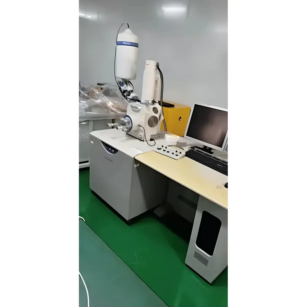
Hitachi S-3400N Used Scanning Electron Microscope (Tungsten-Filament SEM)
| Brand | Hitachi High-Technologies |
|---|---|
| Origin | Japan |
| Model | S-3400N |
| Operating Age | ~10 years |
| Vacuum System | Turbo-molecular pump |
| SE Resolution | 3.0 nm @ 30 kV (high vacuum), 10 nm @ 3 kV (high vacuum) |
| BSE Resolution | 4.0 nm @ 30 kV (low vacuum) |
| Accelerating Voltage | 0.3–30 kV |
| Magnification | ×5 to ×300,000 |
| Max Sample Diameter | 200 mm |
| Stage Types | Manual (Type I) and Motorized 5-Axis (Type II) |
| Stage Travel (Type II) | X 0–100 mm, Y 0–50 mm, Z 5–65 mm, R 360°, T –20° to +90° |
| Max Sample Height | 80 mm (WD = 10 mm) |
| Detector Configuration | High-sensitivity semiconductor BSE detector (5-segment), Everhart-Thornley SE detector |
| Analytical Compatibility | Simultaneous EDX, WDX, and EBSD integration supported |
| Beam Control | Auto filament saturation, auto gun alignment, auto stigmation, auto focus, auto brightness/contrast, manual & automatic beam current adjustment, fixed-ratio & manual & auto 4-quadrant biasing |
| Aperture | Movable 4-position objective aperture |
Overview
The Hitachi S-3400N is a field-proven, tungsten-filament scanning electron microscope engineered for high-reliability routine imaging and materials characterization in academic, industrial QA/QC, and failure analysis laboratories. Based on Hitachi High-Technologies’ established S-series platform, the S-3400N employs thermionic emission from a robust tungsten hairpin filament and operates across a broad accelerating voltage range (0.3–30 kV), enabling versatile contrast optimization for conductive, semi-conductive, and lightly coated specimens. Its dual-mode vacuum architecture—supporting both high-vacuum (HV) and low-vacuum (LV) operation—facilitates stable imaging of non-conductive or hydrated samples without extensive metallization. With secondary electron (SE) resolution of 3.0 nm at 30 kV (HV mode) and 10 nm at 3 kV (HV mode), and backscattered electron (BSE) resolution of 4.0 nm at 30 kV (LV mode), the system delivers reproducible topographic and compositional contrast suitable for microstructural evaluation, particle sizing, fracture analysis, and cross-sectional metrology. The integrated turbo-molecular pumping system ensures rapid pump-down, low hydrocarbon contamination, and long-term column cleanliness—critical for maintaining consistent signal-to-noise performance over extended operational lifecycles.
Key Features
- Automated beam optimization suite: fully integrated software routines for auto filament saturation, auto gun alignment, auto stigmation, auto focus, auto brightness/contrast, and dynamic beam current control
- High-sensitivity 5-segment semiconductor BSE detector enabling quantitative atomic number (Z)-contrast mapping and crystallographic orientation correlation
- Dual-stage configuration: Type I (manual mechanical stage) and Type II (motorized 5-axis stage with ±20° to +90° tilt, 360° rotation, and extended Z travel up to 65 mm)
- Expandable analytical chamber: standardized ports and clearance accommodate simultaneous installation of energy-dispersive X-ray spectroscopy (EDS/EDX), wavelength-dispersive X-ray spectroscopy (WDS/WDX), and electron backscatter diffraction (EBSD) detectors
- Movable 4-position objective aperture for rapid optimization of depth of field, resolution, and signal intensity based on working distance and magnification
- Fixed-ratio, manual, and auto 4-quadrant biasing modes for adaptive SE/BSE signal separation and artifact suppression
Sample Compatibility & Compliance
The S-3400N accommodates specimens up to 200 mm in diameter and 80 mm in height (Type II stage, WD = 10 mm), supporting bulk metals, ceramics, geological thin sections, polymer composites, and biological tissue stubs. Low-vacuum capability (6–270 Pa) permits direct observation of uncoated insulators, powders, and hydrated samples—reducing preparation time and mitigating charging artifacts. All vacuum interlocks, emission controls, and high-voltage safety circuits comply with IEC 61010-1:2010 (Safety Requirements for Electrical Equipment for Measurement, Control, and Laboratory Use). While the instrument predates modern digital audit-trail mandates, its hardware architecture supports post-upgrade integration with GLP/GMP-compliant data acquisition systems meeting FDA 21 CFR Part 11 requirements when paired with validated third-party software.
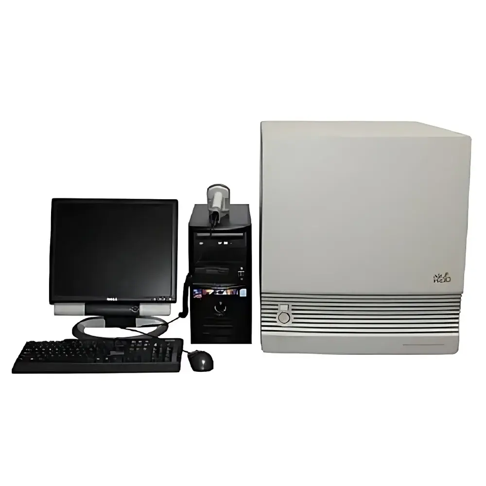
Software & Data Management
Control is executed via dedicated Hitachi PC-based interface using mouse, keyboard, and ergonomic manual knobs—providing deterministic real-time response during manual alignment or fine-tuning. Image acquisition, annotation, measurement (line, area, particle count), and basic histogram analysis are embedded in the native platform. Raw image files (TIFF, BMP) and parameter logs (kV, WD, mag, aperture position, detector bias) are timestamped and exportable for traceability. Although the original software does not natively support cloud synchronization or role-based access control, exported datasets integrate seamlessly with open-format analysis tools including ImageJ/Fiji, MATLAB, and commercial EDS quantification suites (e.g., Oxford AZtec, Thermo Pathfinder).
Applications
- Metallurgical quality control: inclusion identification, grain boundary analysis, weld integrity assessment
- Electronics manufacturing: solder joint inspection, PCB trace delamination, MEMS device morphology
- Geosciences: mineral phase discrimination, porosity quantification, fossil ultrastructure
- Pharmaceutical development: excipient particle morphology, tablet coating uniformity, API crystallinity screening
- Academic research: nanomaterial dispersion analysis, catalyst support topology, biomimetic surface replication
FAQ
Is the S-3400N compatible with modern EDS detectors?
Yes—the sample chamber includes standard flanges and electrical feedthroughs supporting OEM and third-party EDS systems (e.g., Bruker QUANTAX, EDAX Octane). Mechanical and software integration may require vendor-specific interface modules.
What maintenance history is recommended prior to recommissioning?
A full vacuum system validation (leak check, pump oil replacement, turbomolecular pump bearing health assessment), filament replacement, and column cleaning are strongly advised. Calibration of stage encoders and detector gain curves should be performed by qualified service engineers.
Can the system operate under GLP conditions?
While the base hardware lacks built-in electronic signatures or audit trails, it can be operated within GLP frameworks when paired with validated external acquisition software, documented SOPs, and periodic performance qualification (PQ) per ISO/IEC 17025 guidelines.
What is the typical warm-up and stabilization time?
Approximately 30–45 minutes from cold start to thermal and emission stability, assuming ambient lab temperature remains within 20–25°C and humidity <60% RH.
Does the S-3400N support variable pressure imaging?
It supports low-vacuum mode (6–270 Pa) but does not offer true variable pressure (VP-SEM) with differential pumping or gaseous secondary electron detection; imaging of non-conductors relies on charge dissipation via environmental gas ionization rather than signal amplification.
Related Products
")

")
")