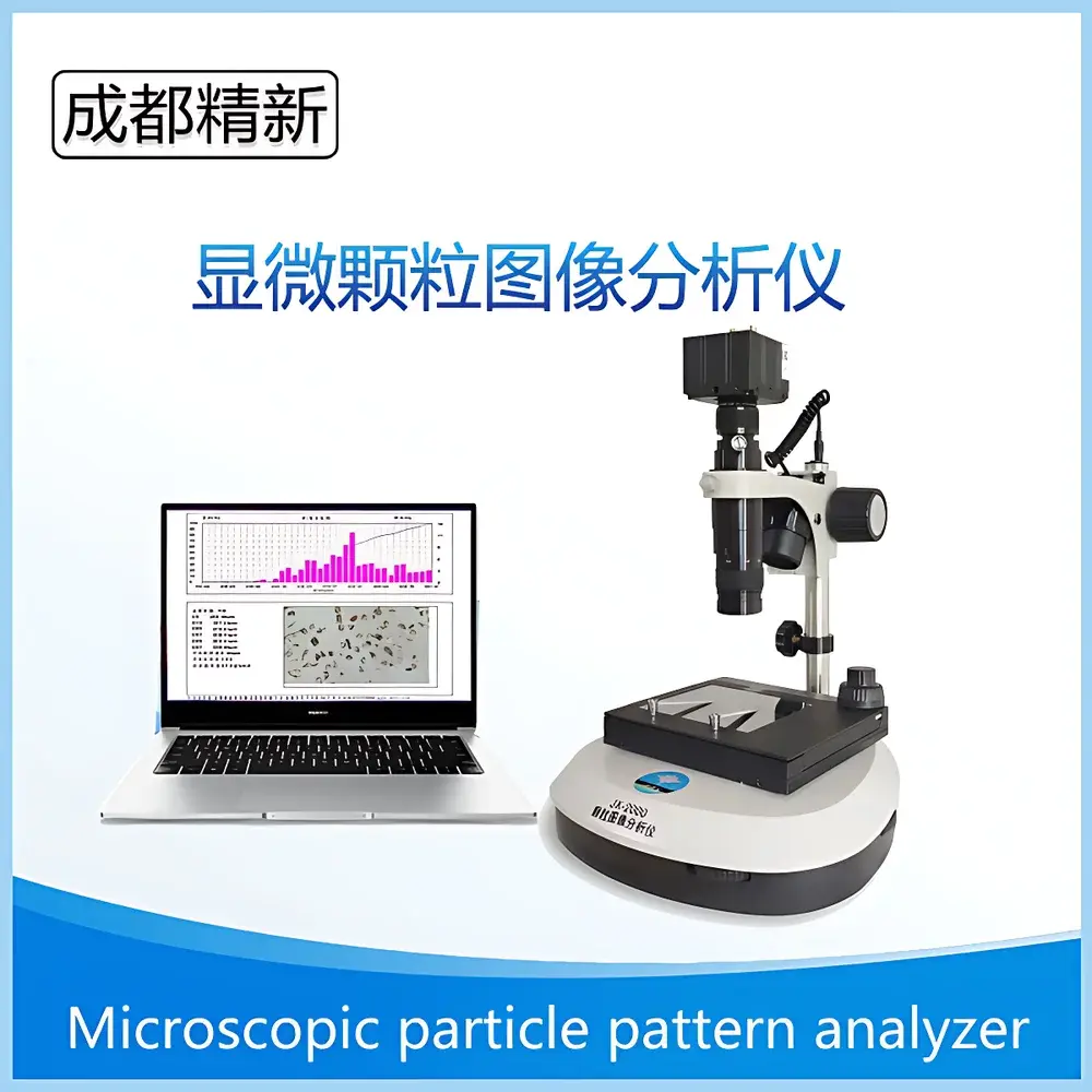
Chengdu Jingxin JX-2000T Microscopic Particle Image Analyzer
| Brand | Chengdu Jingxin (CDJX) |
|---|---|
| Origin | Sichuan, China |
| Manufacturer Type | Direct Manufacturer |
| Regional Classification | Domestic (China) |
| Model | JX-2000T |
| Dispersion Method | Dry & Wet Dispersion |
| Instrument Type | Laboratory-Based Microscopic Image Analyzer |
| Measurement Range | 0.5 µm to 3000 µm |
| Repeatability | < 3% |
| Analysis Time per Sample | ~10 minutes |
| Optical Magnification | Up to 1600× |
| Digital Magnification | Up to 4000× |
| Resolution | ≥ 0.1 µm |
| Microscope Configuration | Transmitted/Reflected Light Microscope (Optional: Domestic, Metallurgical, or Imported Objective Turret) |
| Camera | 3 MP or 5 MP CCD |
| Eyepieces | 10×, 16× |
| Objectives | 4×, 10×, 20×, 40×, 100× |
| Calibration Scale | 10 µm |
| Software Platform | Windows 7 (32-bit) |
| Interface | High-Speed USB |
Overview
The Chengdu Jingxin JX-2000T Microscopic Particle Image Analyzer is a laboratory-grade optical imaging system engineered for quantitative morphological and dimensional characterization of solid particulates in powder, slurry, and suspension forms. Unlike ensemble-averaging techniques such as laser diffraction, this instrument employs direct optical microscopy coupled with high-resolution digital imaging and deterministic image segmentation algorithms to deliver particle-by-particle measurement data—including equivalent circular diameter, Feret length, aspect ratio (length-to-width), circularity, convexity, solidity, and projected area. Its operational principle relies on calibrated brightfield/darkfield transmitted or reflected illumination, precise stage positioning, and pixel-to-micron spatial calibration using traceable NIST-traceable scale bars. Designed for routine QC/QA workflows and R&D validation, the JX-2000T bridges the gap between bulk statistical methods and single-particle metrology—enabling users to correlate physical appearance (e.g., angularity, agglomeration state, surface texture) with functional performance in downstream processes.
Key Features
- Simultaneous acquisition of multiple morphological descriptors per particle: size distribution (D10, D50, D90, D97), aspect ratio distribution, circularity histogram, convexity index, and projected area-weighted statistics.
- Modular microscope platform supporting both transmitted-light (for transparent/semi-transparent particles) and reflected-light (for opaque metals, ceramics, pigments) observation modes; optional upgrade paths to metallurgical or imported objective turrets for enhanced resolution and chromatic correction.
- High-fidelity digital imaging chain: selectable 3 MP or 5 MP progressive-scan CCD camera with low-noise analog-to-digital conversion, synchronized shutter control, and real-time preview at full frame rate.
- Comprehensive image preprocessing suite: grayscale normalization, contrast-limited adaptive histogram equalization (CLAHE), geometric correction (rotation, mirroring, arbitrary scaling), noise suppression (median filtering), and binary threshold optimization (Otsu, IsoData, manual).
- Robust segmentation engine with adaptive edge detection, hole filling, particle splitting/merging logic, and interactive manual editing tools (region erasure, boundary drawing, particle linking) to ensure analytical integrity across heterogeneous samples.
- Batch processing capability for multi-field-of-view (FOV) stitching and statistical aggregation—supporting >10,000 particles per analysis session to meet ISO 13322-1:2020 recommendations for representative sampling.
Sample Compatibility & Compliance
The JX-2000T accommodates dry powders (via static dispersion on conductive stubs or glass slides) and liquid-based suspensions (using standard petri dishes or custom flow cells). It is routinely deployed for quality assessment of abrasives (diamond, silicon carbide), battery cathode materials (LiCoO₂, NMC), catalyst supports (alumina, zeolites), pharmaceutical excipients (lactose, microcrystalline cellulose), food-grade fillers (talc, calcium carbonate), and construction additives (fly ash, silica fume). The system complies with core principles outlined in ISO 13322-1 (Particle Size Analysis — Image Analysis Methods — Part 1: Static Image Analysis) and supports audit-ready documentation required under GLP and GMP environments. All calibration procedures reference traceable 10 µm scale bars; software logs include timestamped operator ID, instrument configuration, and version-controlled analysis parameters—facilitating alignment with FDA 21 CFR Part 11 requirements when paired with validated Windows 7 deployment protocols.
Software & Data Management
The proprietary Jingxin Image Analysis Software (v5.x) operates natively on Windows 7 (32-bit) and communicates with hardware via high-bandwidth USB 2.0 interface. It implements hierarchical data management: raw image files (BMP/JPEG), annotated TIFFs with embedded metadata, and structured XML-based result archives containing full particle tables (X/Y coordinates, major/minor axis, perimeter, area, circularity), summary statistics (mean, std dev, skewness, kurtosis), and customizable report templates. Export options include CSV for third-party statistical packages (e.g., JMP, Minitab), PDF with embedded color thumbnails, and Excel-compatible XLSX with pivot-ready fields. For regulated laboratories, optional configuration enables electronic signature capture, change history tracking, and role-based access control—preparing the system for internal audit readiness and external regulatory review.
Applications
- Verification and cross-validation of laser diffraction or dynamic light scattering results—particularly for non-spherical, anisotropic, or highly polydisperse systems where Mie theory assumptions break down.
- Root-cause analysis of batch-to-batch variability in grinding/milling operations by correlating shape descriptors (e.g., aspect ratio shift toward elongation) with rheological behavior or compaction density.
- Characterization of thermal spray feedstock powders where sphericity and surface roughness directly influence coating porosity and adhesion strength.
- Pharmaceutical granule evaluation per USP guidance—assessing agglomerate integrity, friability-induced fines generation, and blend uniformity via spatial distribution mapping.
- Geotechnical soil particle classification per ASTM D422, supplementing sieve analysis with shape-based sorting indices (e.g., Cz = circularity × sphericity).
FAQ
What sample preparation methods are supported?
Dry dispersion on carbon tape or conductive stubs; wet dispersion in ethanol, isopropanol, or aqueous surfactant solutions using ultrasonic bath or probe sonication (optimized dwell time required per material).
Can the system analyze particles smaller than 0.5 µm?
No—the practical lower limit is constrained by optical diffraction limits and pixel resolution; sub-500 nm features require SEM-based image analysis.
Is automated focus stacking available?
Not natively; focus is manually adjusted per field of view. However, Z-stack acquisition can be implemented via third-party motorized stage integration (custom quotation required).
Does the software support ASTM or ISO-compliant reporting templates?
Yes—preconfigured report layouts align with ISO 13322-1 Annex A and ASTM E2457-17 conventions, including uncertainty estimation notes and method validation references.
How is measurement traceability maintained?
Through daily verification using certified 10 µm scale bars; all calibration events are logged with operator ID, date/time stamp, and deviation values stored within the analysis archive.


