Artinis CD DISC 2.0 Contrast-Detail Phantom for Fluoroscopic Imaging Systems
| Brand | Artinis |
|---|---|
| Country of Origin | Netherlands |
| Model | CD DISC 2.0 |
| Material | Acrylic (PMMA) |
| Tolerance | ±0.03 mm |
| Hole Depth Range | 0.3–8.0 mm (exponential increment) |
| Hole Diameter Range | 0.3–8.0 mm (exponential increment) |
| Total Segments | 240 |
| Concentric Rings | 16 |
| Radial Spokes | 15 |
| Phantom Thickness | Adjustable via stacked acrylic plates |
Overview
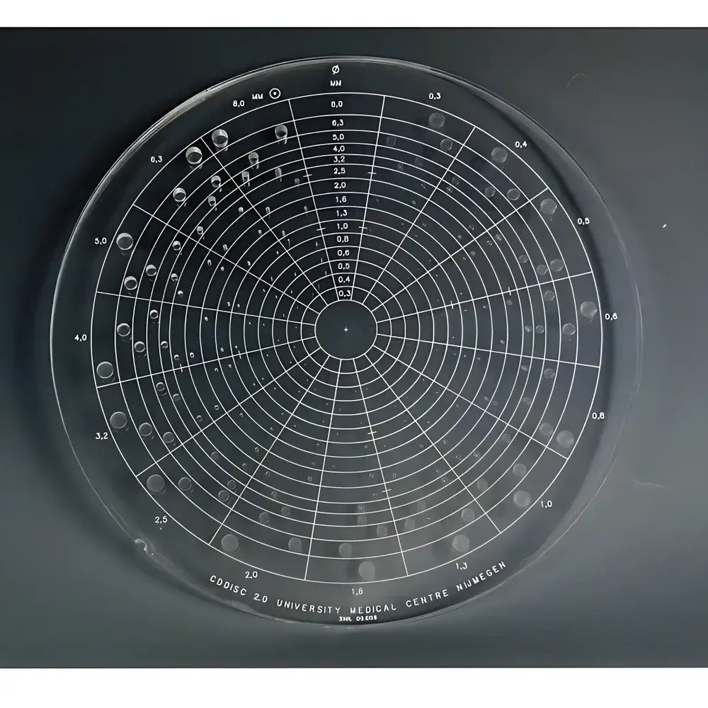
The Artinis CD DISC 2.0 is a precision-engineered contrast-detail (CD) phantom designed specifically for objective, observer-based evaluation of fluoroscopic imaging system performance. Unlike purely physical metrics such as modulation transfer function (MTF) or noise power spectrum (NPS), the CD DISC 2.0 addresses the critical perceptual component of diagnostic imaging—human visual detection thresholds under clinically relevant low-contrast, low-dose conditions. It operates on the principle of psychophysical threshold testing: observers identify the spatial location of subtle high-density spots embedded in a uniform background, enabling quantification of the minimum detectable contrast at varying detail sizes. This dual-parameter assessment—simultaneously mapping contrast sensitivity and spatial resolution—makes the CD DISC 2.0 an essential tool for routine quality control (QC), equipment commissioning, dose optimization studies, and regulatory compliance verification in interventional radiology, cardiac catheterization labs, and neuroradiology suites.
Key Features
- Precision-machined acrylic (PMMA) construction with dimensional tolerances maintained at ±0.03 mm across all cylindrical holes—ensuring metrological traceability and inter-laboratory reproducibility.
- 240 discrete test segments arranged in 16 concentric rings and 15 radial spokes, each containing a single high-contrast spot positioned randomly in one of four corners to eliminate positional bias.
- Exponentially increasing hole depth (0.3 mm → 8.0 mm clockwise per ring) to generate 15 distinct contrast levels; simultaneously, exponentially increasing hole diameter (0.3 mm → 8.0 mm radially outward) to produce 16 graded spatial detail levels.
- Modular design allows stacking of additional PMMA plates to simulate variable patient thicknesses (e.g., 10 cm, 20 cm, 30 cm equivalent attenuation), enabling system evaluation under realistic beam-hardening and scatter conditions.
- Compatible with all fluoroscopic systems—including image intensifier-based, flat-panel detector (FPD), and digital subtraction angiography (DSA) platforms—without requiring proprietary software or calibration routines.
Sample Compatibility & Compliance
The CD DISC 2.0 is validated for use in accordance with international standards governing medical imaging quality assurance, including IEC 62220-1-2 (characterization of digital x-ray detectors), AAPM Report No. 31 (quality control in fluoroscopy), and EN 61223-3-1 (acceptance and constancy testing). Its design reflects the methodology originally established by Thijssen et al. (Neuroradiology, 1988) and Rose (Vision: Human and Electronic, 1974), forming the basis for observer performance testing referenced in FDA guidance documents for fluoroscopic device submissions. The phantom supports GLP-compliant QC protocols when integrated into documented, auditable workflows—particularly where observer variability must be tracked across time, personnel, or equipment upgrades.
Software & Data Management
While the CD DISC 2.0 is a hardware-only phantom and requires no dedicated software, its output is fully compatible with standardized CD curve analysis frameworks. Institutions routinely digitize acquired phantom images using DICOM-compliant viewers (e.g., OsiriX, Horos, or vendor-neutral PACS workstations) and record observer responses via structured spreadsheets or custom web-based scoring interfaces. For regulatory submissions or multi-center trials, data collection may be aligned with 21 CFR Part 11 requirements through electronic signature-enabled platforms that enforce audit trails, user authentication, and version-controlled reporting. The phantom’s deterministic geometry ensures that raw observer data—binary “detected/not detected” per segment—can be directly plotted as contrast-detail curves using open-source tools (e.g., Python SciPy, R psychometric packages) or commercial QA software suites (e.g., QAPLUS, Image Owl).
Applications
- Baseline characterization and longitudinal monitoring of fluoroscopic system performance during preventive maintenance cycles.
- Dose reduction validation—quantifying trade-offs between reduced mAs/kVp settings and maintained observer detection capability.
- Comparative evaluation of detector technologies (e.g., CsI vs. a-Se FPDs) under identical acquisition parameters.
- Observer training and competency assessment for radiologists, interventional cardiologists, and radiologic technologists.
- Supporting ISO 9001 and ACR Accreditation documentation for medical physics QA programs.
- Research applications in visual psychophysics, human factors engineering, and AI-assisted detection algorithm benchmarking.
FAQ
Is the CD DISC 2.0 suitable for CT or mammography systems?
No—it is optimized exclusively for fluoroscopic and DSA imaging geometries, with geometry, contrast range, and spatial scale calibrated to typical kVp (60–120 kV), pulse rates (1–30 fps), and detector entrance doses encountered in real-time x-ray imaging.
Can multiple observers be evaluated simultaneously using the same image?
Yes. Standardized viewing conditions (controlled ambient lighting, fixed monitor luminance, calibrated display gamma) enable concurrent independent scoring, supporting inter-observer agreement analysis (e.g., Cohen’s kappa, intraclass correlation).
Does Artinis provide certified calibration documentation?
The CD DISC 2.0 is supplied with a manufacturer’s dimensional certificate verifying hole depth and diameter tolerances (±0.03 mm); however, it is not a metrology standard traceable to NIST or EURAMET—users are advised to verify geometric fidelity annually using optical coordinate measuring systems.
How often should CD testing be performed?
Per AAPM recommendations, CD phantom evaluation should be conducted at installation, after major service events, and at least quarterly as part of routine fluoroscopy QC programs.
Is there a digital version or automated scoring algorithm available?
No official automated scoring software is distributed by Artinis. Automated detection algorithms remain unsuitable for CD analysis due to fundamental differences between machine vision and human contrast sensitivity—observer-based scoring remains the clinical gold standard per current regulatory consensus.