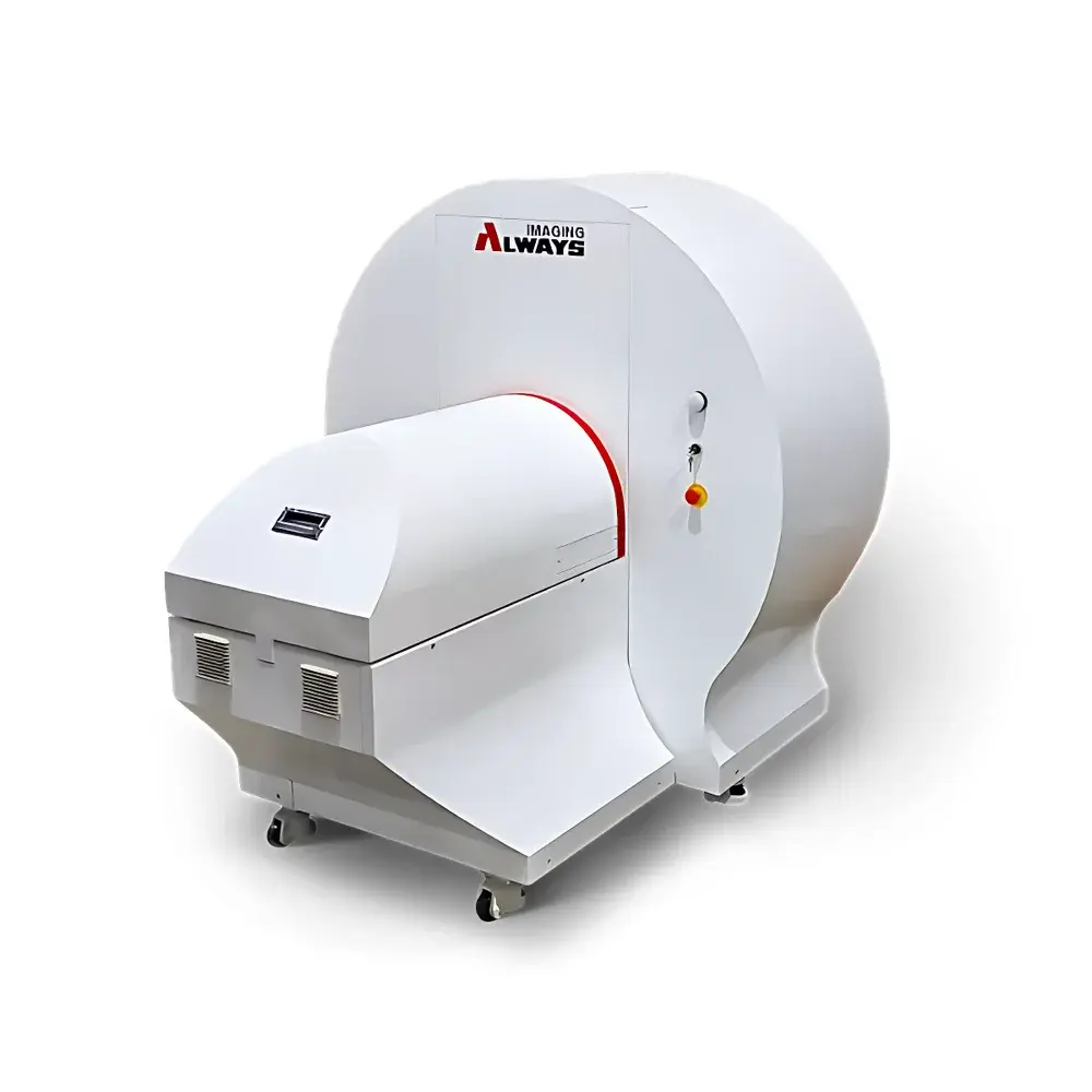
Always Imaging AX-Life 2000CT High-Resolution In Vivo and Ex Vivo Small Animal Computed Tomography System
| Brand | Always Imaging |
|---|---|
| Origin | Jiangsu, China |
| Manufacturer Type | Manufacturer |
| Origin Category | Domestic |
| Model | AX-Life 2000CT |
| Price | Upon Request |
| Spatial Resolution | 4 µm |
Overview
The Always Imaging AX-Life 2000CT is a benchtop high-resolution micro-computed tomography (micro-CT) system engineered for quantitative 3D morphological and functional analysis of small animal models—both in vivo and ex vivo. Operating on the principle of cone-beam X-ray computed tomography, the system acquires projection data through rotational acquisition around the specimen axis, followed by iterative or analytical reconstruction into isotropic volumetric datasets. Designed specifically for preclinical research laboratories, the AX-Life 2000CT delivers sub-5 µm spatial resolution without compromising scan throughput or dose efficiency—enabling longitudinal studies with high anatomical fidelity and reproducible quantitative metrics across timepoints.
Key Features
- Sub-5 µm isotropic spatial resolution (best achievable 4 µm), validated per ASTM E1441 Annex A3 protocols using calibrated line-pair phantoms;
- Continuous variable geometric magnification via precise source-to-object-to-detector positioning, eliminating fixed-magnification constraints;
- Dual-acquisition modes: step-and-shoot (for high-stability static imaging) and continuous gantry rotation (enabled by slip-ring connectivity), supporting scan durations as short as 3.9 seconds per full 360° rotation;
- High-fidelity detector array: 2340 × 2882 pixels, 50 µm pitch, 16-bit dynamic range, coupled with air-cooled, integrated microfocus X-ray source (5 µm focal spot, 20–100 kV adjustable, max 15 W output);
- Proprietary spatial beam shaping technology reduces effective radiation dose by 2–3× relative to conventional collimation—validated against ISO 4037-3 reference dosimetry—while preserving contrast-to-noise ratio (CNR) and modulation transfer function (MTF) performance;
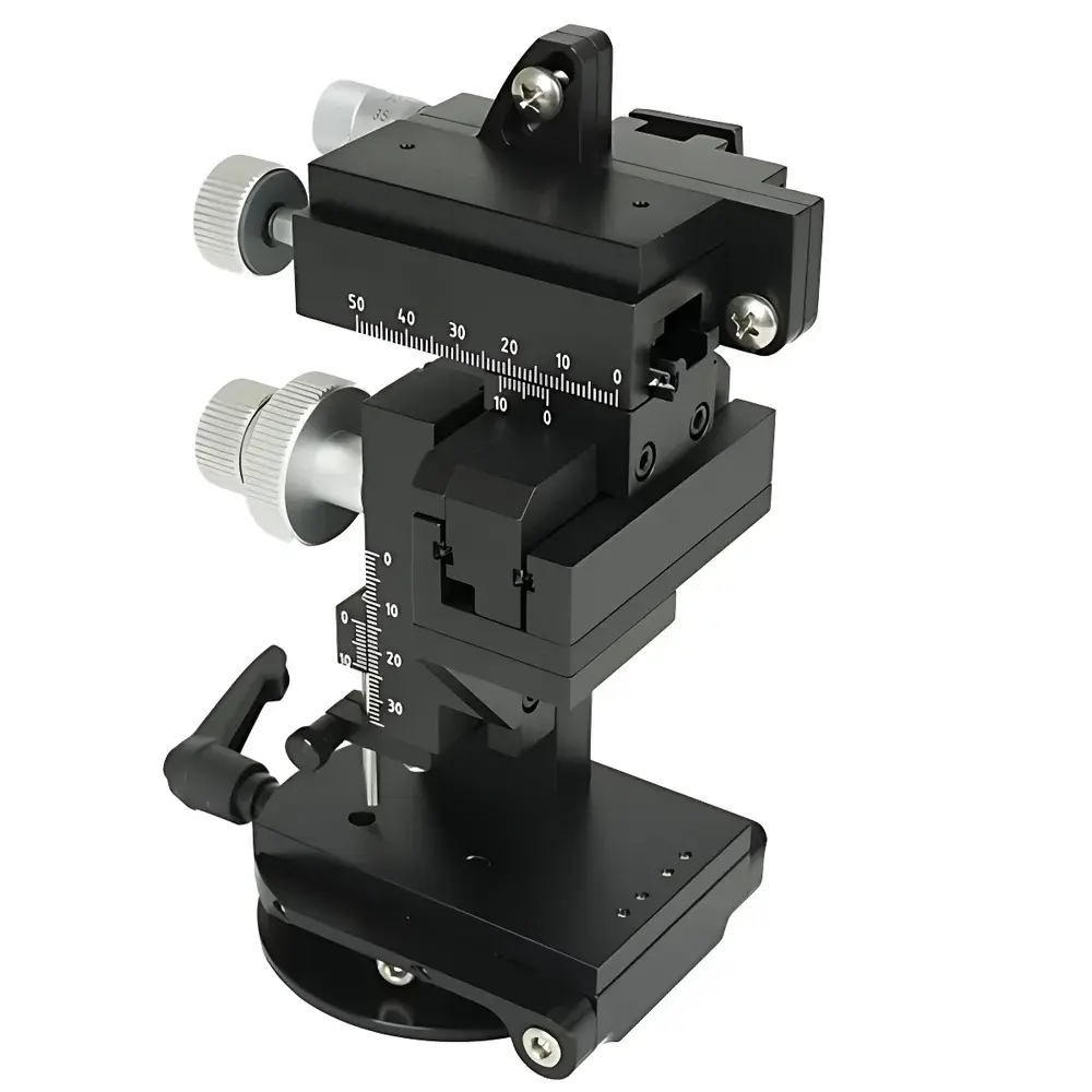
- Modular animal handling platform: quick-swap mouse and rat beds with integrated physiological monitoring ports (optional), rated for up to 5 kg payload and compatible with stereotactic frames;
- GPU-accelerated reconstruction engine integrating InstaRecon® hierarchical algorithm architecture, delivering >10× faster reconstruction versus standard Feldkamp-Davis-Kress (FDK) implementations on identical hardware.
Sample Compatibility & Compliance
The AX-Life 2000CT accommodates a broad spectrum of biological specimens—from live anesthetized rodents (C57BL/6, Sprague-Dawley, nude mice) to excised tissues (bone, lung, vasculature, tumor xenografts), mineralized scaffolds, and biomaterial implants. All scanning protocols adhere to ALARA (As Low As Reasonably Achievable) radiation safety principles. The system meets IEC 62494-1:2017 requirements for preclinical X-ray imaging devices and complies with national Class II radiation equipment regulations (CNCA-C11-01:2023). Shielding integrity is certified to deliver <1 µSv/h at 5 cm from any external surface under maximum operational conditions (per GBZ 138–2022).
Software & Data Management
The unified acquisition and reconstruction suite provides intuitive, workflow-driven operation—including one-click scan initiation, real-time preview reconstruction, and automated beam hardening correction. DICOM 3.0 export is natively supported for PACS integration. Reconstruction outputs are stored in standardized NIfTI-1 format with embedded metadata (voxel size, exposure time, kV/mA, reconstruction kernel). Audit trails comply with GLP and 21 CFR Part 11 requirements when deployed with optional electronic signature modules; all user actions, parameter changes, and reconstruction logs are timestamped and immutable.
Applications
- Longitudinal bone microarchitecture analysis (trabecular thickness, separation, BV/TV) per ASBMR guidelines;
- Pulmonary airspace quantification in COPD and fibrosis models;
- Vascular casting and angiographic phenotyping (e.g., tumor neovascularization, cerebral collateral circulation);
- Implant-bone interface assessment in orthopedic and dental biomaterial studies;
- Ex vivo organ phenotyping (kidney glomeruli, pancreatic islets, myocardial infarct volume);
- Material science applications: pore network modeling in hydrogels, scaffold porosity grading, and additive manufacturing QA.
FAQ
What is the minimum achievable voxel size in vivo?
The system achieves isotropic voxels down to 4 µm in optimized ex vivo scans; in vivo applications typically operate between 8–20 µm depending on motion control, dose constraints, and subject size.
Is the system compatible with respiratory and cardiac gating?
Yes—external TTL-compatible physiological signal inputs (via BNC) enable retrospective sorting and prospective triggering for thoracic and cardiac imaging.
Can raw projection data be exported for third-party reconstruction?
Yes—unprocessed sinogram data (in HDF5 format) and geometry metadata are fully accessible via software SDK for custom algorithm development.
Does the system support multi-energy or spectral CT capabilities?
Not natively; however, dual-kV sequential scanning and post-hoc material decomposition workflows are supported through optional software modules.
What regulatory documentation is provided for institutional biosafety and radiation safety committees?
Full technical dossier—including shielding test reports, dose calibration certificates, IEC compliance summaries, and risk analysis per ISO 14971—is supplied with each installation.