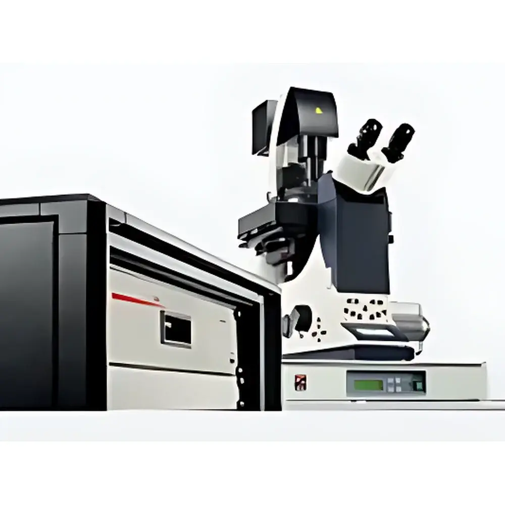
Leica SR GSD 3D Super-Resolution Laser Microdissection System
| Brand | Leica |
|---|---|
| Origin | Germany |
| Model | Leica SR GSD 3D |
| Imaging Principle | Ground-State Depletion (GSD) / direct Stochastic Optical Reconstruction Microscopy (dSTORM) |
| Lateral Resolution | ≤20 nm |
| Axial Resolution | ≤50 nm |
| Optical Configuration | Automated Total Internal Reflection Fluorescence (TIRF) |
| Objective Correction | Apochromatic |
| Calibration Options | Gold nanoparticle reference, fluorescent bead-based, dye-specific spectral calibration |
| Software | Leica GSD 3D Acquisition & Analysis Suite (compliant with GLP audit trails and ISO/IEC 17025 traceability workflows) |
Overview
The Leica SR GSD 3D is a fully integrated super-resolution laser microdissection platform engineered for nanoscale spatial mapping and precise subcellular isolation in three dimensions. Unlike conventional widefield or confocal systems, it combines ground-state depletion (GSD) and direct stochastic optical reconstruction microscopy (dSTORM) modalities within a rigorously stabilized TIRF architecture. This enables deterministic single-molecule localization with quantifiable uncertainty—critical for correlating molecular identity, spatial distribution, and functional context in fixed and live-cell specimens. The system operates on the physical principle of photoswitchable fluorophore control: by transiently depleting the ground state population via targeted laser excitation, it achieves sparse, temporally resolved emission events that are computationally reconstructed into a diffraction-unlimited image stack. Its axial resolution of ≤50 nm—achieved through engineered astigmatism correction and multi-plane PSF fitting—represents a fundamental advancement over standard widefield dSTORM platforms, which typically exhibit axial localization precision >300 nm.
Key Features
- Automated, motorized TIRF illumination with real-time angle optimization for consistent evanescent field depth control across heterogeneous sample topographies
- Dual-channel, high-quantum-efficiency sCMOS detection with synchronized laser gating (405 nm activation, 640 nm readout, optional 561 nm co-excitation)
- Integrated piezo-driven Z-stage with closed-loop feedback (±1 nm repeatability) enabling precise 3D point-spread function (PSF) sampling and drift correction
- Apochromatically corrected optics across 400–800 nm spectrum, minimizing chromatic aberration for multicolor single-molecule co-localization
- On-the-fly drift compensation using fiducial gold nanoparticles (20–40 nm) embedded in coverslip substrate or co-immobilized with biological samples
- Modular design supporting optional integration with Leica DMi8 inverted microscope base, environmental chamber (37°C/5% CO₂), and motorized XY stage for large-area tile scanning
Sample Compatibility & Compliance
The Leica SR GSD 3D accommodates standard No. 1.5H glass coverslips (24 × 60 mm or 22 × 22 mm), custom-patterned substrates, and polymer-based cell culture inserts compatible with long-term live imaging. It supports immunolabeled, genetically encoded (e.g., mMaple, PAmCherry), and self-labeling (HaloTag/SNAP-tag) probes. All optical pathways conform to DIN EN ISO 10110-5 standards for surface quality and wavefront error (<λ/10 RMS). The system meets IEC 61000-6-3 (EMC emissions) and IEC 61000-6-2 (immunity) requirements. Data acquisition and processing modules comply with ALCOA+ principles; audit trail functionality, electronic signatures, and version-controlled protocol storage satisfy FDA 21 CFR Part 11 and EU Annex 11 regulatory expectations for regulated research environments.
Software & Data Management
Leica GSD 3D Acquisition & Analysis Suite provides end-to-end workflow management—from raw photon stream capture to 3D localization rendering and quantitative colocalization analysis. Key modules include: (1) Real-time localization engine with maximum-likelihood estimation (MLE) and Bayesian uncertainty propagation; (2) Multi-color registration using iterative closest point (ICP) algorithms with sub-pixel affine correction; (3) Volumetric ROI definition for downstream laser microdissection targeting; (4) Export to standardized formats (OME-TIFF, HDF5) with embedded metadata per OME-XML schema. All calibration routines—including gold nanoparticle reference mapping and dye-specific PSF library generation—are traceable to NIST-traceable standards and logged with timestamped operator credentials.
Applications
- Quantitative mapping of synaptic protein nanodomains in neuronal tissue sections
- 3D reconstruction of nuclear pore complex architecture and transport receptor occupancy
- Spatial profiling of chromatin accessibility markers (e.g., ATAC-seq targets) with sub-100 nm positional fidelity
- Correlative super-resolution imaging and laser-capture microdissection (LCM) of morphologically defined subcellular compartments
- Validation of CRISPR-Cas9 editing efficiency via simultaneous visualization of guide RNA scaffold and target locus
- Time-resolved tracking of endocytic vesicle maturation using dual-color dSTORM with <100 ms temporal binning
FAQ
What distinguishes Leica SR GSD 3D from conventional STORM or PALM systems?
It integrates hardware-optimized TIRF with computational astigmatism engineering and closed-loop Z-control—enabling isotropic 3D localization without requiring multi-plane acquisition or post-hoc deconvolution approximations.
Can the system perform live-cell super-resolution imaging?
Yes—when paired with environmental control and low-phototoxicity dyes (e.g., Alexa Fluor 647 with oxygen scavenging systems), it supports time-lapse dSTORM at frame rates up to 2 Hz with maintained ≤50 nm axial precision.
Is calibration data exportable for third-party analysis?
All calibration parameters—including PSF models, drift trajectories, and channel registration matrices—are exportable in HDF5 format with documented units and coordinate conventions.
Does the software support batch processing of multi-condition datasets?
Yes—the Analysis Suite includes scriptable pipelines (Python API) for automated localization, clustering (DBSCAN), and statistical comparison across experimental replicates under user-defined significance thresholds.
How is instrument performance validated post-installation?
Leica provides on-site verification using NIST-traceable fluorescent nanobead standards (100 nm diameter), with full report documenting lateral/axial FWHM, localization precision (σxy/σz), and photobleaching kinetics per channel.


