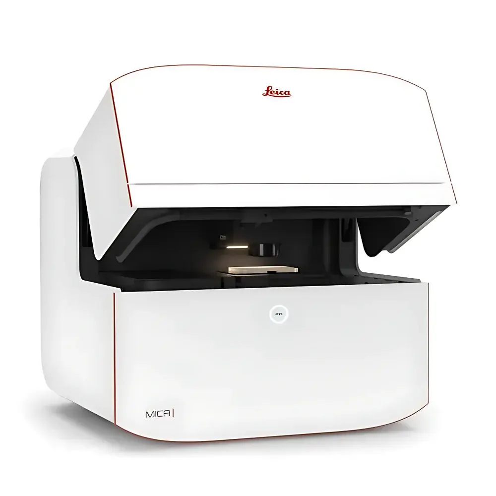
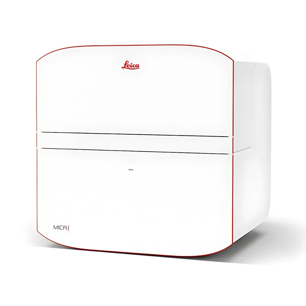
Leica MICA Widefield-Confocal Multimodal Imaging Analysis Platform
| Brand | Leica |
|---|---|
| Origin | Germany |
| Manufacturer Type | Authorized Distributor |
| Origin Category | Imported |
| Model | MICA Widefield-Confocal |
| Instrument Type | Point-Scanning Confocal Microscope |
| Lasers | 405 nm, 488 nm, 561 nm, 638 nm |
| Fluorescence Detectors | Standard configuration with four HyD detectors and four sCMOS cameras |
| Optical Zoom | 1–6× |
| Microscope Host | Fully motorized inverted fluorescence microscope |
| Illumination Sources | Laser + LED |
| Software & Workstation | Intuitive GUI with integrated Pixel Classifier AI module |
| Vibration Isolation | Passive anti-vibration table |
Overview
The Leica MICA Widefield-Confocal Multimodal Imaging Analysis Platform represents a paradigm shift in optical microscopy—redefining accessibility, reproducibility, and analytical depth for both expert and non-specialist users. Engineered around a unified point-scanning confocal architecture combined with high-sensitivity widefield imaging, MICA enables true spatiotemporal correlation across up to four fluorescent labels in a single acquisition. Unlike conventional sequential multi-channel imaging—where sample drift, stage repositioning, or temporal delays compromise registration—MICA’s FluoSync spectral unmixing technology synchronizes excitation and detection pathways across all lasers and detectors, delivering inherently registered 4-channel data with sub-pixel spatial fidelity and millisecond-level temporal alignment. This is achieved through hardware-integrated spectral decomposition and real-time signal routing, eliminating the need for post-acquisition alignment or channel interpolation. The platform integrates a fully motorized inverted microscope chassis, environmental chamber (temperature, CO₂, humidity control), and intelligent optical path management—making it equally suitable for fixed-tissue analysis, live-cell dynamics, and long-term 3D organoid monitoring.
Key Features
- Fully automated multimodal acquisition: OneTouch optimization adjusts illumination intensity, detector gain, pinhole size, z-step interval, and exposure time based on user-defined priority—from “sample preservation” to “maximum resolution.”
- Simultaneous widefield and confocal imaging: Captures complementary structural (widefield) and optical-sectioned (confocal) data within the same field of view and timepoint, enabling correlative context without registration artifacts.
- FluoSync spectral separation: Patented hardware-software co-design allows concurrent excitation and detection of up to four fluorophores (e.g., DAPI, FITC, Cy3, Cy5) with minimal crosstalk and zero temporal offset.
- AI-powered Pixel Classifier: Trains on user-drawn annotations directly within the GUI; generates reusable, project-specific segmentation models compliant with GLP documentation requirements (audit trail enabled).
- THUNDER and LIGHTNING computational imaging modules: THUNDER delivers deconvolution-based background suppression for thick specimens; LIGHTNING applies adaptive structure-enhancement algorithms optimized for subcellular features—both embedded natively in the acquisition workflow.
- Integrated live-cell incubation chamber: Maintains physiological conditions (37°C, 5% CO₂, >95% RH) over multi-day acquisitions; minimizes medium evaporation via sealed chamber design and active humidity regulation.
Sample Compatibility & Compliance
MICA supports diverse biological specimens—including adherent and suspension cells, tissue sections (frozen and FFPE), 3D spheroids, organoids, and microtiter plate assays (up to 384-well format). Its dual-mode capability ensures optimal contrast for both thin monolayers (via confocal optical sectioning) and thick volumetric samples (via widefield + THUNDER processing). All system components comply with IEC 61000-6-3 (EMC) and IEC 61000-6-2 (immunity) standards. The software architecture conforms to FDA 21 CFR Part 11 requirements for electronic records and signatures, including role-based access control, full audit trail logging, and electronic signature validation. Data export formats (OME-TIFF, ND2, CZI) adhere to FAIR principles and are compatible with downstream analysis pipelines certified under ISO/IEC 17025-accredited laboratories.
Software & Data Management
Leica Application Suite X (LAS X) MICA Edition provides an integrated environment for acquisition, visualization, AI training, and quantitative analysis. The GUI features contextual toolbars that adapt dynamically to selected modality (widefield/confocal), objective, and fluorophore set. All imaging parameters—including laser power, HyD voltage, camera binning, and z-stack settings—are stored as metadata within OME-TIFF files. The Pixel Classifier module supports supervised learning with cross-validation metrics (Dice coefficient, precision/recall) and exports trained models as portable .pkl files for inter-laboratory deployment. Raw and processed datasets can be archived in hierarchical folder structures synchronized with institutional LIMS via configurable network paths. Audit logs record every parameter change, user login, model training event, and image export—fully traceable for regulatory submissions.
Applications
- High-content screening: Simultaneous 4-color imaging of apoptosis markers (Caspase-3/7, mitochondrial membrane potential, actin cytoskeleton, nuclear morphology) in 96-well plates with <10 µm well-to-well positional repeatability.
- 3D tissue phenotyping: Correlative widefield overview (20×) and confocal zoom (63× water immersion) of intestinal crypts, enabling seamless transition from architectural assessment to subcellular protein localization (e.g., detyrosinated tubulin networks).
- Long-term live-cell dynamics: 72-hour timelapse of GFP-tagged MX1 expression in MDCK spheroids at 30-min intervals under controlled CO₂ and temperature—preserving viability while capturing growth kinetics and protein trafficking.
- Neuroscience workflows: Multiplexed staining of rat brain sections (DAPI, FITC-STL, Cy3-GFAP, Cy5-NeuN) with automatic focus map generation (Sample Finder) and region-of-interest selection prior to high-resolution confocal acquisition.
- Quantitative organelle analysis: AI-driven segmentation of mitochondria (MitoTracker Green/TMRE) coupled with morphometric quantification (aspect ratio, form factor, network connectivity) using built-in measurement tools calibrated per objective magnification.
FAQ
What distinguishes MICA from conventional confocal systems?
MICA eliminates sequential channel acquisition through hardware-synchronized FluoSync detection, ensuring absolute spatiotemporal correlation across all four channels—critical for kinetic studies and multiplexed biomarker quantification.
Can MICA perform true optical sectioning in widefield mode?
No—widefield mode captures full-depth projection; however, THUNDER computational clearing provides near-confocal contrast for thick samples without physical sectioning or increased phototoxicity.
Is the Pixel Classifier module validated for regulated environments?
Yes—the module includes IQ/OQ documentation templates, version-controlled model repositories, and full audit trail export compliant with GxP and ISO 13485 requirements.
Does MICA support third-party objective lenses?
Only Leica-certified objectives with encoded mechanical and optical parameters are supported to ensure accurate magnification calibration and automatic correction of spherical/chromatic aberrations.
How is data integrity maintained during extended timelapse experiments?
The system performs real-time checksum verification of each acquired frame, logs thermal drift compensation events, and triggers alerts if environmental parameters deviate beyond ±0.3°C or ±0.2% CO₂ from setpoints.


