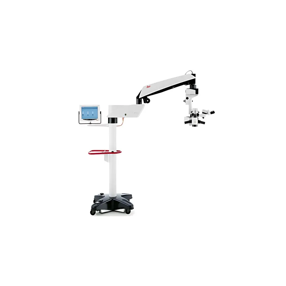
Leica M844 F40 / F20 Ophthalmic Surgical Microscope
| Brand | Leica |
|---|---|
| Country of Origin | Germany |
| Model | M844 F40 / F20 |
| Optical System | APO OptiChrome |
| Illumination | Direct Halogen with OttoFlex Independent Red Reflex Illumination |
| Zoom Technology | QuadZoom Synchronized Quad-Optical Path |
| Assistant Scope | Dual-Arm Stereo Assistant Microscope |
| Imaging | HD Video Recording & Real-Time Monitor Output |
| Control Interface | Touchscreen Display with One-Touch Video Mode |
Overview
The Leica M844 F40 / F20 Ophthalmic Surgical Microscope is an advanced, CE-marked surgical visualization platform engineered for precision anterior and posterior segment procedures—including cataract extraction, corneal transplantation, glaucoma filtration surgery, vitreoretinal interventions, and refractive lens exchange. Built upon Leica’s legacy in high-fidelity optical engineering, the M844 integrates APO OptiChrome achromatic-apochromatic optics to deliver diffraction-limited resolution, minimal chromatic aberration, and exceptional color fidelity across the visible spectrum (400–700 nm). Its core optical architecture employs a dual-path illumination design: direct halogen illumination ensures uniform, shadow-free tissue illumination at clinically safe irradiance levels (<15 mW/cm² at the cornea), while the integrated OttoFlex red reflex illumination system provides independent, adjustable 635 nm red light—critical for intraoperative assessment of lens capsule integrity, capsulorhexis centration, and vitreous cavity visualization without increasing overall photic load. The microscope operates on a stable, vibration-damped mechanical base with motorized focus and zoom drives compliant with ISO 13485:2016 and IEC 60601-1 safety standards.
Key Features
- QuadZoom synchronized quad-optical path technology enables simultaneous, parfocal magnification adjustment for both primary surgeon and assistant scopes—eliminating manual re-focusing during collaborative procedures.
- Dual-arm stereo assistant microscope with independent interpupillary distance adjustment (55–75 mm) and ergonomic articulation supports teaching, proctoring, and multi-operator workflows in academic medical centers.
- High-efficiency APO OptiChrome optical train achieves >92% light transmission efficiency—reducing required illumination power by up to 40% compared to conventional xenon-based systems, thereby minimizing thermal load and phototoxic risk to ocular tissues.
- Integrated touchscreen control panel with one-touch video mode toggles the display between real-time surgical feed, parameter configuration, and playback of HD (1080p/60 fps) recorded procedures—fully compatible with DICOM-SR structured reporting workflows.
- Motorized zoom (F40: 6.3×–40×; F20: 4×–20×), continuous focus range (±12 mm), and programmable preset recall support standardized setup protocols aligned with GLP-compliant surgical documentation requirements.
Sample Compatibility & Compliance
The M844 F40 / F20 is validated for use with human cadaveric eyes, porcine ex vivo models, and live animal ophthalmic models (e.g., rabbit, non-human primate) under IACUC-approved protocols. It complies with essential regulatory frameworks including EU MDR 2017/745 Annex I (General Safety and Performance Requirements), IEC 62304 (medical device software lifecycle), and FDA 21 CFR Part 11 (electronic records and signatures) when paired with Leica’s certified surgical documentation software suite. All optical components are autoclavable or compatible with hospital-grade disinfectants (e.g., 70% isopropyl alcohol, hydrogen peroxide vapor), meeting EN 14885 standards for reusable medical device reprocessing.
Software & Data Management
Leica’s proprietary SURGICAL SUITE software (v4.2+) provides secure, audit-trail-enabled video capture, timestamped metadata logging (magnification, illumination intensity, zoom position), and encrypted export to PACS or EMR systems via HL7/FHIR interfaces. Built-in DICOM connectivity supports integration into enterprise imaging infrastructures compliant with IHE Eye Care Technical Framework profiles. Video recordings include embedded calibration markers traceable to NIST-traceable reference standards, satisfying ISO/IEC 17025 requirements for measurement uncertainty documentation in clinical research settings.
Applications
- Cataract surgery: Capsulorhexis guidance, phacoemulsification monitoring, IOL positioning verification
- Corneal surgery: DALK, DSEK, DMEK graft orientation and interface assessment
- Vitreoretinal surgery: Membrane peeling, endolaser application, silicone oil–air interface visualization
- Glaucoma surgery: Trabeculectomy bleb formation, gonioscopy-assisted transluminal trabeculotomy (GATT)
- Neuro-ophthalmic procedures: Optic nerve head dissection, orbital tumor resection
- Preclinical research: In vivo murine retinal imaging, zebrafish optic tectum mapping, and ex vivo human donor eye histopathology correlation studies
FAQ
Is the M844 compatible with third-party surgical navigation systems?
Yes—the microscope features a standardized VESA mount and RS-232/USB-C API interface for bidirectional communication with platforms such as Zeiss CALLISTO eye, Alcon Verion, and Topcon INTEGRAL.
What is the maximum working distance for the F40 and F20 configurations?
F40: 200 mm ±5 mm; F20: 250 mm ±5 mm—both configurable with optional extended working distance objectives (up to 300 mm) per ISO 10940-1.
Does the system support FDA 21 CFR Part 11 compliance for electronic signatures?
When deployed with Leica SURGICAL SUITE v4.2+ and validated IT infrastructure, full Part 11 compliance—including role-based access control, electronic signature audit trails, and record retention policies—is achievable.
Can the OttoFlex red reflex illumination be calibrated independently of main illumination?
Yes—OttoFlex output is digitally regulated via closed-loop photodiode feedback, allowing independent intensity adjustment from 0–100% in 1% increments without affecting main beam spectral balance or thermal stability.


