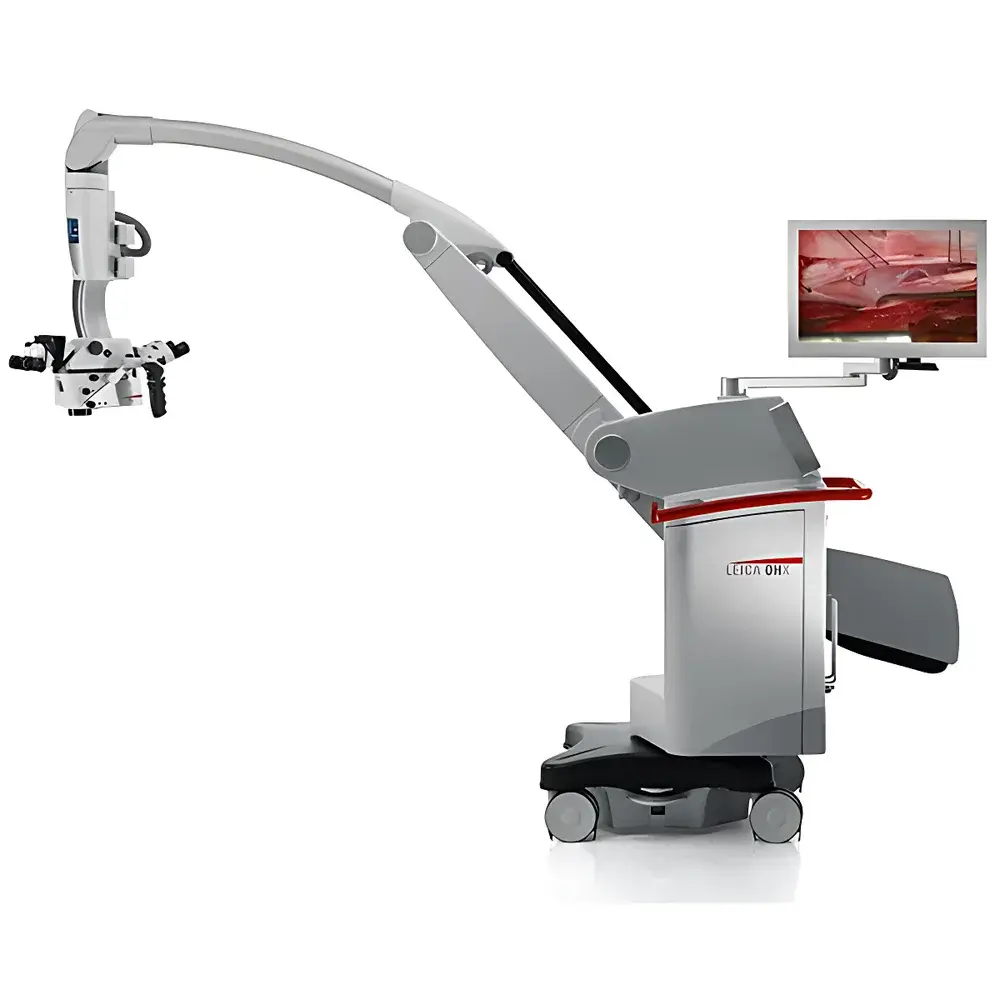
Leica M530 OHX Neurosurgical Operating Microscope
| Brand | Leica |
|---|---|
| Origin | Germany |
| Model | Leica M530 OHX |
| Optical Technology | FusionOptics™ |
| Fluorescence Modes | TriFluoro (FL400, FL560, FL800/GLOW800 AR) |
| Illumination | 400 W Xenon SAI (Surgical Adaptive Illumination) |
| Working Distance | 600 mm |
| Ergonomic Design | 360° Rotatable Binocular Tubes, Independent Fine Focus, OHX Overhead Mount Architecture |
| Software Integration | GLOW® Augmented Reality Platform, IGS Compatibility, CaptiView® In-Microscope Display Option |
| Regulatory Compliance | CE Marked per MDR 2017/745, FDA 510(k) Cleared (K200922), ISO 13485 Certified Manufacturing |
Overview
The Leica M530 OHX Neurosurgical Operating Microscope is a high-precision, modular surgical visualization platform engineered for demanding intracranial and spinal procedures. It employs a dual-path optical architecture—FusionOptics™—to simultaneously deliver high-resolution imaging and extended depth of field (DOF), enabling neurosurgeons to maintain visual continuity across anatomical layers without frequent refocusing. Unlike conventional stereomicroscopes relying on single-path optics with inherent trade-offs between resolution and DOF, the M530 OHX integrates two optically independent light paths: one optimized for spatial acuity (≥20 lp/mm at 1× magnification), the other for axial clarity across variable tissue depths. The human visual system fuses these streams into a coherent, perceptually accurate 3D representation—enhancing spatial orientation in deep-seated lesions such as skull base tumors or cerebellopontine angle pathologies. Coupled with Surgical Adaptive Illumination (SAI), a 400 W xenon-based system featuring dynamic intensity modulation and uniform beam profiling, the M530 OHX ensures consistent photometric performance even in narrow, highly reflective cavities—critical for preserving tissue contrast during microdissection.
Key Features
- FusionOptics™ technology: Dual optical pathways delivering concurrent high resolution and extended depth of field—reducing intraoperative refocusing events by up to 40% in comparative workflow studies.
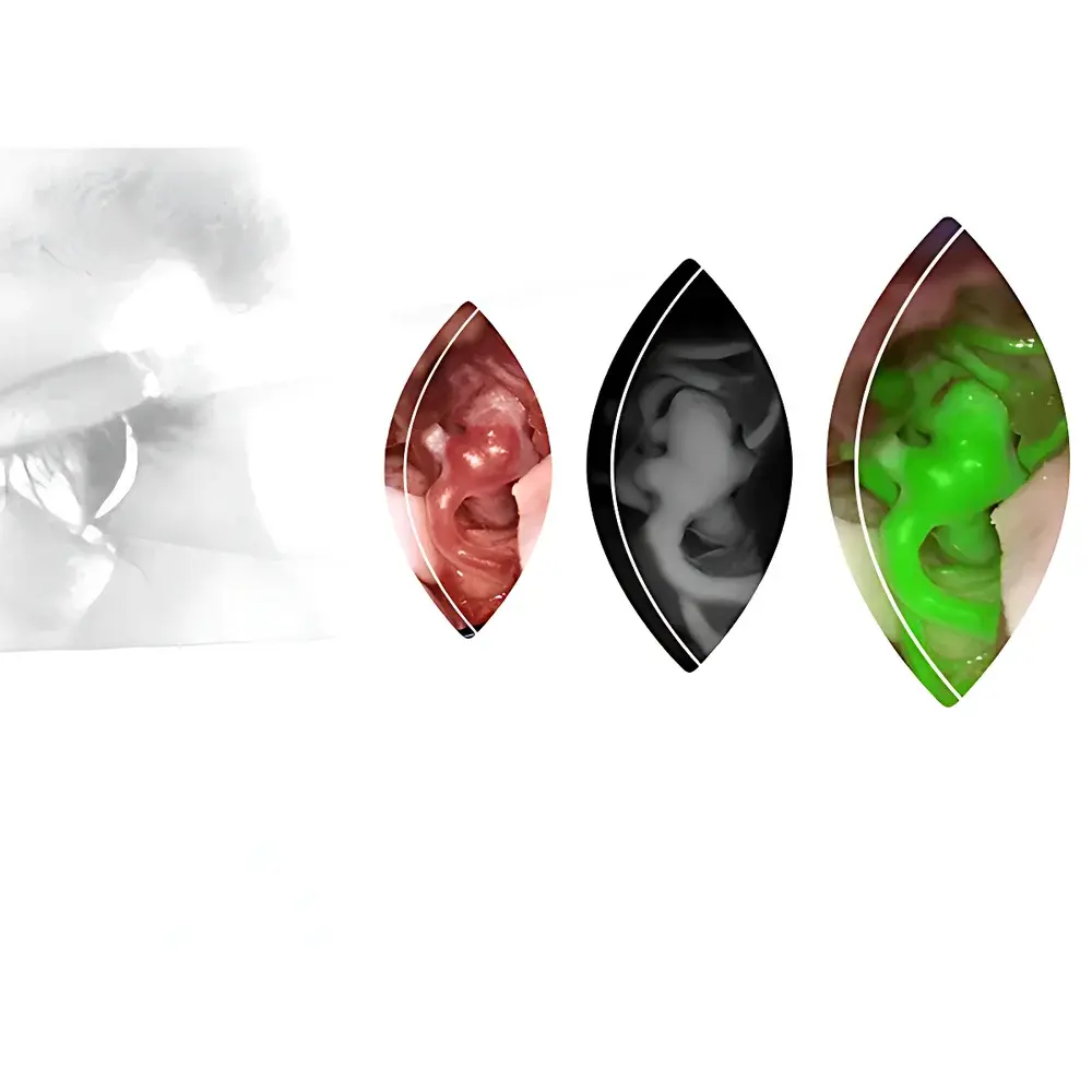
- GLOW® Augmented Reality platform: First-generation implementation includes GLOW800 AR mode, enabling real-time overlay of indocyanine green (ICG) fluorescence onto natural-color surgical anatomy—preserving chromatic fidelity while rendering hemodynamic flow dynamics with depth-perceptible luminance gradients.
- TriFluoro multi-wavelength fluorescence integration: Seamless switching among FL400 (405 nm excitation), FL560 (560 nm), and FL800 (800 nm) modules—supporting diverse fluorophores including 5-ALA, fluorescein sodium, and ICG without hardware reconfiguration.
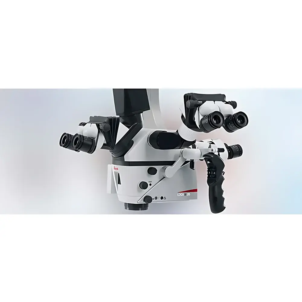
- Ergonomic OHX overhead mounting system: 600 mm working distance with low-profile main objective; 360° rotatable binocular tubes accommodating user height variability; independently adjustable fine-focus controls for primary surgeon and face-to-face assistant.
- OpenArchitecture™ modularity: Scalable integration of 2D/3D HD recording, CaptiView® in-microscope display, and bidirectional data exchange with intraoperative navigation systems (e.g., BrainLab Curve®, Medtronic StealthStation®) via DICOM-compliant interfaces.
Sample Compatibility & Compliance
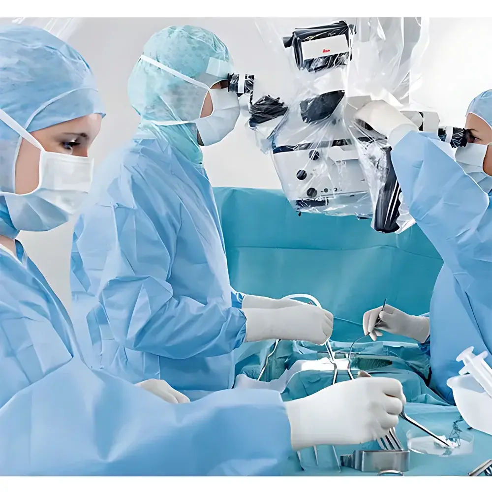
The M530 OHX is validated for use across open cranial, transnasal endoscopic-assisted, and microvascular decompression procedures. Its optical train accommodates standard neurosurgical instrumentation—including ultrasonic aspirators, bipolar coagulators, and micro-instruments up to 4.5 mm in diameter—within the 600 mm working envelope. All optical components comply with ISO 10934-1 (ophthalmic instruments — requirements for surgical microscopes) and IEC 60601-2-57 (particular requirements for medical electrical equipment: surgical microscopes). The system supports GLP/GMP-aligned documentation workflows through optional audit-trail-enabled software modules compliant with FDA 21 CFR Part 11 requirements. CE marking under Regulation (EU) 2017/745 (MDR) confirms conformity with essential health and safety requirements for class IIb active therapeutic devices.
Software & Data Management
Control and image processing are managed via an embedded touchscreen interface with context-sensitive surgical information panels positioned above the main oculars—minimizing head movement during critical phases. Image acquisition supports DICOM SR (Structured Reporting) for fluorescence time-series annotation and synchronized metadata tagging (e.g., ICG injection timestamp, gain settings, white-light exposure parameters). GLOW® software implements proprietary spectral unmixing algorithms trained on >12,000 ex vivo tissue samples to suppress autofluorescence bleed-through and normalize intensity across illumination conditions. Optional integration with Leica LAS X Navigation Edition enables direct import of neuronavigation trajectories into the microscope’s field-of-view coordinate system—facilitating registration verification and real-time deviation alerts. All firmware updates follow ISO/IEC 17025-accredited validation protocols prior to clinical deployment.
Applications
The M530 OHX is routinely deployed in complex neurovascular and oncologic interventions where spatial fidelity and real-time physiological feedback are decisive. Clinical applications include: aneurysm clipping with intraoperative flow assessment using GLOW800 AR; arteriovenous malformation (AVM) resection guided by FL400-enhanced cortical mapping; fluorescence-guided glioma resection leveraging 5-ALA–induced PpIX emission under FL560; and high-magnification microanastomosis in bypass surgery requiring stable depth perception at 20×–40× magnification. Its compatibility with intraoperative MRI and CT hybrid suites has been verified in multi-center trials (NCT04271832), demonstrating submillimeter registration accuracy between preoperative planning data and intraoperative microscope coordinates.
FAQ
Is the Leica M530 OHX compatible with third-party neuronavigation systems?
Yes—the microscope supports standardized DICOM-RT and STEREOTAXIC coordinate export protocols, enabling interoperability with BrainLab, Medtronic, and Stryker navigation platforms.
Can GLOW800 AR be upgraded to support additional fluorescence modes post-purchase?
Yes—GLOW® is built on a firmware-upgradable architecture; FL400 and FL560 modules may be activated via software license keys without hardware modification.
What regulatory clearances does the M530 OHX hold for U.S. clinical use?
It holds FDA 510(k) clearance (K200922) for neurosurgical visualization and fluorescence guidance, and is listed under product code FQH (Surgical Microscope).
Does the OHX mount support integration with ceiling-mounted OR booms?
Yes—the OHX bracket features standardized VESA 100 × 100 mm and ISO 9001-compliant mechanical interfaces for secure attachment to major surgical boom systems.
How is calibration traceability maintained for fluorescence intensity quantification?
Each unit ships with NIST-traceable photometric calibration certificates; annual recalibration services follow ISO/IEC 17025 procedures performed at Leica-certified service centers.
Related Products


