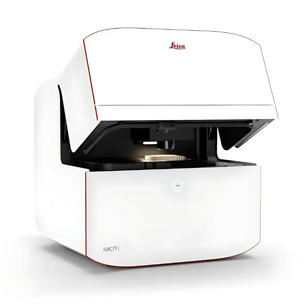
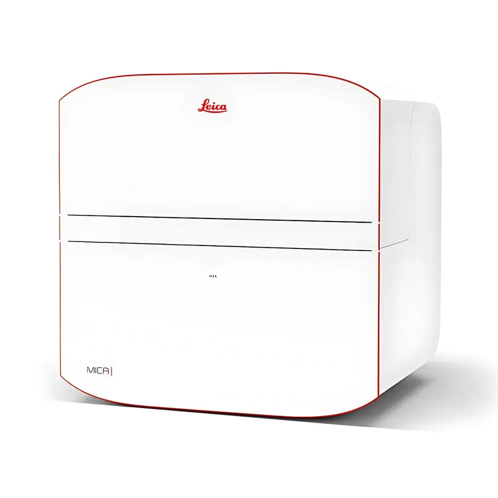
Leica MICA Widefield Live-Cell Multimodal Imaging & Analysis Platform
| Brand | Leica |
|---|---|
| Country of Origin | Germany |
| Instrument Type | Inverted Fluorescence Microscope |
| Excitation Source | LED |
| Objective Options | 63× Water Immersion Objective with Auto-Watering |
| Fluorescence Detection | FluoSync Hyperspectral Unmixing Technology |
| Automation Level | Fully Motorized |
| Focusing Strategy | Three Intelligent Focus Modes (Adaptive, Continuous, and Z-Stack) |
| Observation Interface | Integrated Scientific CMOS Camera |
| DIC Compatibility | Compatible with Glass and Plastic Culture Vessels |
| Stage | Fully Motorized Scanning Stage |
| Filter System | FluoSync Hyperspectral Unmixing for Simultaneous Multi-Channel Acquisition |
Overview
The Leica MICA Widefield Live-Cell Multimodal Imaging & Analysis Platform is an integrated, fully automated inverted fluorescence microscope engineered for high-content, time-resolved analysis of living biological specimens under physiologically relevant conditions. Built upon a modular optical architecture, MICA combines widefield epifluorescence, confocal point-scanning, transmitted light contrast (including DIC), and intelligent environmental control within a single instrument platform. Its core measurement principle relies on simultaneous spectral acquisition via FluoSync — a hardware-software co-optimized hyperspectral unmixing method that captures full emission spectra from up to four fluorophores in a single exposure, eliminating temporal and spatial misregistration inherent in sequential channel acquisition. This enables true spatiotemporal correlation across multiplexed labels — critical for dynamic studies of organelle interactions, cell migration, apoptosis kinetics, and 3D tissue morphogenesis.
Key Features
- Fully motorized optical train with intelligent focus management: Adaptive focus tracking, continuous focus maintenance, and programmable Z-stack acquisition ensure consistent focal plane retention across long-term live-cell experiments.
- FluoSync hyperspectral unmixing engine: Integrates custom dichroic optics, tunable emission filters, and real-time spectral deconvolution algorithms to resolve spectrally overlapping dyes without sequential scanning or spectral crosstalk compensation.
- Integrated environmental chamber: Precisely regulates temperature (±0.2 °C), CO2 (±0.1%), and humidity to sustain viability during extended timelapse imaging — compatible with multiwell plates, Petri dishes, and microfluidic chambers.
- OneTouch workflow automation: Context-aware parameter optimization adjusts illumination intensity, exposure time, gain, and z-step size based on selected imaging priority — ranging from “Sample Protection” (low phototoxicity) to “Image Quality” (maximum SNR).
- Unified multimodal acquisition: Seamlessly switches between widefield, confocal, DIC, and phase contrast modes within the same session — all controlled through a single software interface with synchronized metadata tagging.
- AI-powered image analysis pipeline: Includes pre-trained and user-customizable pixel classifiers for organelle segmentation (e.g., mitochondria, nuclei, actin), integrated THUNDER computational clearing for thick-sample deconvolution, and LIGHTNING sub-diffraction enhancement for ultrastructural detail.
Sample Compatibility & Compliance
MICA supports standard life science consumables including glass-bottom dishes, polymer-based culture plates (e.g., µClear, ibidi), and organoid-compatible scaffolds. Its DIC module is validated for use with both glass and plastic substrates, ensuring compatibility with high-throughput screening workflows. The system complies with ISO 13485 design controls for medical device-related research instrumentation and meets electromagnetic compatibility (EMC) requirements per IEC 61326-1. All digital image acquisition and storage modules adhere to FDA 21 CFR Part 11 guidelines for electronic records and signatures when deployed in GLP/GMP-regulated environments. Audit trails, user access logs, and version-controlled AI model training histories are natively supported.
Software & Data Management
Acquisition and analysis are unified under Leica Application Suite X (LAS X) v4.13+, which provides a structured project-based file architecture compliant with OME-TIFF and HDF5 standards. Raw spectral data cubes (λ–x–y–z–t) are stored alongside processed channels, enabling retrospective re-analysis and spectral re-unmixing. The software includes built-in support for FAIR data principles: automatic metadata embedding (including objective ID, excitation wavelength, detector settings, environmental parameters), batch processing pipelines, and export to common formats (NIS-Elements ND2, Imaris IMS, Bio-Formats). For regulated environments, LAS X supports role-based user permissions, electronic signatures, and full audit trail generation for all acquisition, annotation, and analysis events.
Applications
- Live-cell multiplexed timelapse: Simultaneous monitoring of mitochondrial membrane potential (TMRE), cytoskeletal dynamics (SiR-Actin), caspase activation (CellEvent Caspase-3/7), and nuclear morphology (DAPI) over >72 h at 30-min intervals — with zero registration drift.
- 3D tissue section imaging: Correlative widefield overview (20×) and confocal zoom (63× water) of intestinal crypts, with THUNDER deconvolution applied to remove out-of-focus blur and LIGHTNING enhancement revealing microtubule detyrosination patterns.
- Organoid and spheroid growth quantification: Automated detection and volumetric tracking of GFP-tagged MDCK spheroids cultured in 96-well plates, with environmental stability maintained throughout 60+ hour acquisitions.
- High-content screening (HCS): Full-spectrum acquisition from 4-color fluorescent assays in multiwell formats, eliminating well-to-well illumination variability through OneTouch calibration and auto-exposure normalization.
- AI-augmented phenotypic profiling: User-guided training of pixel classifiers directly on acquired images — e.g., distinguishing apoptotic vs. necrotic cells based on combined TMRE/DAPI/Caspase signal topology — with model portability across instruments and users.
FAQ
Does MICA require external lasers or mercury/xenon lamps?
No. MICA uses solid-state LED illumination across UV–NIR wavelengths (365–770 nm), providing stable, low-heat output with no warm-up time or alignment maintenance.
Can FluoSync be used with conventional filter sets?
FluoSync requires its dedicated spectral detection path and is not compatible with standard bandpass filter cubes; however, legacy filter-based acquisition remains available as a separate mode.
Is the environmental chamber compatible with gas-permeable membranes?
Yes — the chamber accommodates oxygen-permeable materials such as PDMS and specialized cell culture inserts, with optional O2 sensor integration for hypoxia studies.
How is data integrity ensured during long-term acquisitions?
All acquisitions include checksum validation, automatic backup to network-attached storage (NAS), and real-time disk space monitoring with predictive failure alerts.
Can MICA integrate with third-party analysis platforms like ImageJ/Fiji or Python-based tools?
Yes — raw OME-TIFF exports preserve full spectral and metadata fidelity, and LAS X provides RESTful API access for programmatic control and data ingestion into custom analysis pipelines.


