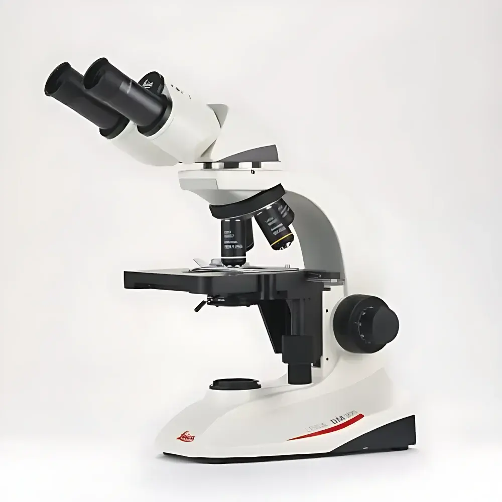
Leica DM300 Teaching Microscope
| Brand | Leica |
|---|---|
| Origin | Shanghai, China |
| Manufacturer Type | Authorized Distributor |
| Country of Origin | China |
| Model | DM300 |
| Price Range | USD 1,400 – 7,200 |
Overview
The Leica DM300 is a robust, entry-level compound microscope engineered specifically for undergraduate life science education. Designed in accordance with Leica Microsystems’ long-standing optical and mechanical heritage—spanning over 170 years—the DM300 delivers clinical-grade optical performance and ergonomic reliability in an education-optimized platform. It operates on standard brightfield microscopy principles, utilizing Köhler illumination geometry to ensure uniform, glare-free specimen illumination across all magnifications. Its optical path supports standard 10× and 40× objective lenses (with optional 100× oil immersion), and is fully compatible with standard 23 mm eyepiece tubes and 22 mm field number eyepieces. The instrument is not intended for fluorescence, phase contrast, or DIC applications; rather, it focuses on delivering high-fidelity, reproducible transmitted-light imaging for histology, cytology, microbiology, and basic botany laboratories.
Key Features
- Compact, space-efficient chassis with integrated base design—occupies less than 0.03 m² footprint, ideal for shared lab benches and mobile teaching carts.
- Sealed, maintenance-free coaxial focusing mechanism featuring hardened brass gears and precision-ground lead screws, ensuring consistent parfocal accuracy and tactile repeatability over >50,000 focusing cycles.
- Leica EZLite™ LED illumination system: cold-white (6,000 K) output with intensity control, rated for ≥20,000 hours MTBF, consuming ≤3.2 W—reducing energy use by ~80% compared to halogen-based predecessors.
- Abbe condenser (NA 1.25) with engraved scale and centering screws, pre-aligned for rapid Köhler setup; includes iris diaphragm with graduated aperture control.
- Low-position mechanical stage with dual-axis vernier controls (X/Y resolution: 0.1 mm), enabling stable, vibration-dampened specimen navigation at 400× and 1000× total magnification.
- Interchangeable observation head options: fixed monocular, rotating monocular (for shared viewing), or Siedentopf binocular tube (100% light split, interpupillary adjustment 55–75 mm).
Sample Compatibility & Compliance
The DM300 accommodates standard 1″ × 3″ glass microscope slides (76 × 26 mm) and 22 × 22 mm coverslips. It supports routine mounting media including glycerol, DPX, and aqueous solutions, and is compatible with common staining protocols (H&E, Gram, Giemsa, Toluidine Blue). All optical components meet ISO 8578:2017 (Microscopes — Requirements for compound microscopes) and conform to IEC 61000-6-3:2019 for electromagnetic compatibility. While not certified for GMP or GLP environments, its mechanical stability, traceable calibration pathways, and documented service history support compliance with institutional QA/QC requirements for academic laboratory accreditation (e.g., ISO/IEC 17025:2017 Annex A.3 for educational instrumentation).
Software & Data Management
The DM300 is a standalone optical instrument with no embedded digital imaging module. However, it features standardized C-mount (23.2 mm) and trinocular port options (optional accessory), enabling seamless integration with third-party USB 3.0 CMOS cameras (e.g., Leica DMC series, AmScope MU1400, or Thorlabs CS2100M). When paired with Leica Application Suite (LAS) X LE or open-source platforms such as Fiji/ImageJ, the system supports annotation, measurement (pixel-to-µm calibration via stage micrometer), time-lapse capture, and export in TIFF, PNG, or JPEG formats. Audit trails, user access logs, and electronic record retention are managed externally through institutional LIMS or learning management systems (LMS); the microscope itself does not store data or require FDA 21 CFR Part 11-compliant software.
Applications
- Undergraduate histology labs: identification of epithelial, connective, muscular, and nervous tissue architecture.
- Microbiology practicums: bacterial morphology assessment, Gram-stain interpretation, and yeast/bacterial colony isolation verification.
- Botany coursework: stomatal density quantification, vascular bundle analysis in monocot/dicot stems, and pollen grain morphology.
- Cell biology instruction: mitotic stage identification in onion root tip squashes, organelle visualization using vital dyes (e.g., Janus Green B for mitochondria).
- Clinical lab science training: peripheral blood smear evaluation, urinalysis sediment examination, and basic parasitology screening (e.g., Entamoeba histolytica cysts).
FAQ
Is the Leica DM300 suitable for advanced research applications?
No—it is purpose-built for pedagogical use and lacks modular expansion capabilities for advanced contrast techniques or motorized automation.
Can the DM300 be upgraded with phase contrast or fluorescence?
No; its optical train and illumination pathway are fixed for brightfield only. Upgrades require a higher-tier platform such as the Leica DM500 or DM750.
What is the warranty coverage for academic institutions?
Leica Microsystems provides a standard 2-year limited warranty on parts and labor, extendable to 3 years under institutional education service agreements.
Does the DM300 comply with CE or UL safety standards?
Yes—the instrument carries CE marking per Directive 2014/30/EU (EMC) and 2014/35/EU (LVD), and meets UL 61010-1:2012 for laboratory electrical equipment.
Are replacement bulbs required for the LED illumination system?
No—EZLite™ LEDs are permanently mounted and non-replaceable; their rated lifetime exceeds 20 years under typical daily classroom usage (≤4 hrs/day).