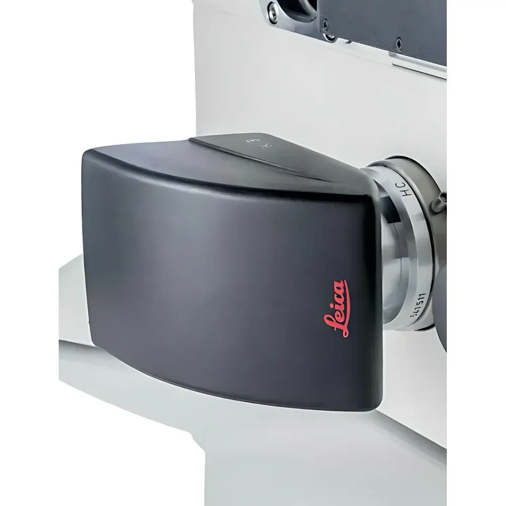
Leica K3 C/M Series Microscope Cameras
| Brand | Leica |
|---|---|
| Origin | Germany |
| Model | K3 C/M |
| Sensor Type | CMOS |
| Color/monochrome variants | K3 C (color), K3 M (monochrome) |
| Bit Depth | 12-bit per channel (RGB) |
| Dynamic Range | High (>70 dB typical) |
| Frame Rate | Up to 60 fps at full resolution |
| Interface | USB 3.0 |
| Software Compatibility | Leica LAS X, Steel/Cleanliness Expert, TWAIN-compliant applications |
| Compliance | CE, RoHS, ISO 13485 (for clinical use), FDA 21 CFR Part 11 ready (with LAS X audit trail) |
Overview
The Leica K3 C/M Series Microscope Cameras are high-performance digital imaging solutions engineered for precision, reproducibility, and workflow efficiency across life science research, industrial quality control, and clinical diagnostics. Built on modern CMOS sensor architecture, the K3 platform delivers quantitative imaging capabilities grounded in photometric accuracy and low-noise signal acquisition. Unlike legacy CCD-based systems, the K3 leverages global shutter CMOS technology to eliminate motion artifacts during rapid scanning or time-lapse acquisition—critical for high-throughput tissue screening, metallurgical cleanliness analysis, and forensic evidence documentation. Its optical design is optimized for integration with Leica upright and inverted microscopes, including widefield systems equipped with motorized stages and automated illumination. The camera’s native 12-bit RGB output ensures linear response across intensity gradients, enabling reliable pixel-level quantification required for fluorescence intensity mapping, spectral unmixing (when paired with filter sets), and morphometric analysis.
Key Features
- High-fidelity 12-bit RGB image capture with calibrated color reproduction—essential for histopathology, immunofluorescence, and multi-channel co-localization studies.
- Wide dynamic range (>70 dB) supports simultaneous visualization of weak and saturated signals in heterogeneous fluorescent samples—e.g., low-expression membrane proteins alongside bright nuclear DAPI staining.
- USB 3.0 interface enables sustained data throughput up to 60 fps at native resolution, significantly reducing mosaic acquisition time for large-area scans compared to prior-generation cameras.
- Dual-variant architecture: K3 C (color) for true-color documentation and phenotypic assessment; K3 M (monochrome) for maximum quantum efficiency in low-light fluorescence or brightfield contrast techniques such as phase contrast and DIC.
- TWAIN driver support ensures seamless interoperability with third-party image analysis platforms—including MATLAB-based custom pipelines, HALO®, Visiopharm®, and LIS/HIS/LIMS environments compliant with IHE PDI or HL7 standards.
- Robust mechanical housing rated IP54 for dust and splash resistance—suitable for shared core facilities and regulated production environments.
Sample Compatibility & Compliance
The K3 C/M series accommodates diverse specimen types without optical recalibration: stained tissue sections (H&E, Masson’s trichrome), fluorescently labeled cell cultures (GFP/RFP/mCherry), metallic surface contaminants (ISO 4406 particle counting), forensic trace evidence (hair, fiber, gunshot residue), and semiconductor wafer defects. It meets ISO/IEC 17025 requirements for measurement uncertainty reporting when used with NIST-traceable calibration targets. For clinical deployment, the system aligns with EN ISO 15189:2022 (medical laboratories) and supports GLP/GMP audit trails via LAS X Navigator’s electronic logbook—recording operator ID, timestamp, exposure parameters, and post-acquisition processing steps. All firmware updates undergo formal change control per IEC 62304 Class B software lifecycle management.
Software & Data Management
Leica LAS X Navigator serves as the primary acquisition and stitching engine, offering automated Z-stack alignment, multi-position mosaic generation, and batch export in TIFF, JPEG2000, or OME-TIFF formats compliant with Bio-Formats. The Steel/Cleanliness Expert module provides ASTM E2812-compliant particle classification and size distribution reporting for metallurgical applications. Raw K3 data retains embedded EXIF metadata (exposure time, gain, white balance, lens ID), ensuring traceability under FDA 21 CFR Part 11 when configured with digital signature and role-based access controls. Exported datasets integrate directly into LIMS via RESTful API or DICOM-SR wrappers for PACS archival.
Applications
- Life Sciences: High-content screening of organoid cultures, mitotic index quantification in tumor biopsies, and spatial transcriptomics slide validation.
- Industrial Metrology: ISO 13322-2-compliant particle morphology analysis on steel surfaces; coating thickness uniformity assessment via reflectance imaging.
- Clinical Pathology: Digital pathology slide digitization at 20×–40× magnification with color fidelity validated against College of American Pathologists (CAP) benchmarks.
- Forensics: Side-by-side comparison of questioned documents using multispectral reflectance ratios captured under controlled LED illumination sequences.
- Materials Science: In-situ corrosion monitoring via time-lapse brightfield imaging synchronized with environmental chamber telemetry.
FAQ
Is the K3 C/M compatible with non-Leica microscope stands?
Yes—via C-mount adapter and third-party drivers—but optimal performance (including auto-exposure synchronization and hardware-triggered acquisition) requires Leica DM-series or ICC50-compatible stands.
Does the K3 support live streaming to remote collaboration platforms?
Yes—LAS X includes WebShare functionality enabling real-time browser-based viewing with adjustable compression profiles and session-based user permissions.
Can raw K3 images be processed in open-source tools like Fiji/ImageJ?
Yes—TIFF exports retain full bit depth and metadata; Bio-Formats plugin enables direct import with channel registration and scale bar preservation.
What is the recommended maintenance interval for sensor calibration?
Annual verification using Leica’s certified flat-field reference target (P/N 11590127) is advised for GxP-regulated workflows; routine dark-frame subtraction is performed automatically during acquisition.
How does the K3 handle mixed illumination modes (e.g., fluorescence + transmitted light)?
LAS X allows concurrent dual-channel capture with independent exposure control per lamp source, preserving photometric linearity across modalities without manual gain adjustment.


