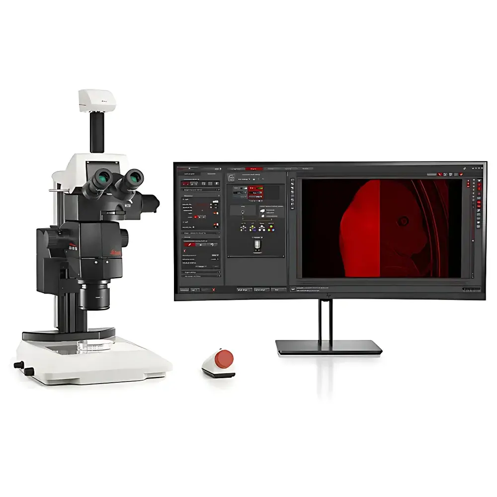
Leica THUNDER Imager Model Organism Automated Macro Fluorescence Microscopy System
| Brand | Leica |
|---|---|
| Origin | Germany |
| Instrument Type | Research-Grade Inverted/ Upright Integrated Fluorescence Microscope |
| Excitation Source | High-Power LED |
| Configuration | Fully Automated Whole-Organism Imaging Platform with Computational Clearing |
| Optical Architecture | Stereo + Epifluorescence + Z-stack Acquisition Engine |
| Compliance | Designed for ISO/IEC 2382:2015-compliant image clarity and structural fidelity in biological imaging |
Overview
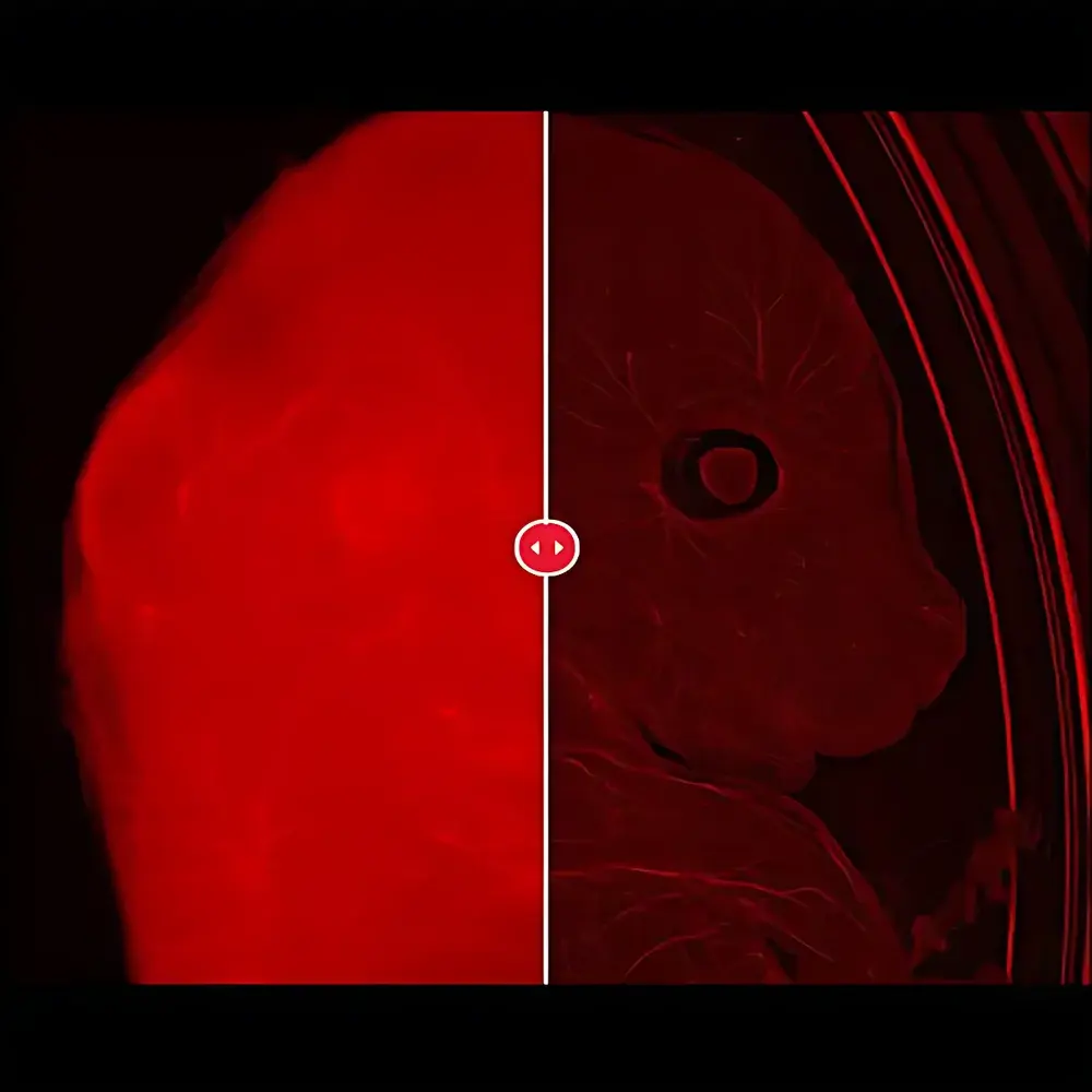
The Leica THUNDER Imager Model Organism is an automated, high-throughput macro fluorescence microscopy system engineered for quantitative 3D exploration of intact living organisms—from Caenorhabditis elegans and Drosophila melanogaster to zebrafish larvae, plant seedlings, and cleared mouse embryos. Unlike conventional widefield systems limited by out-of-focus blur in thick specimens, this platform integrates Leica’s proprietary Instant Computational Clearing (ICC) technology directly into the optical acquisition pipeline. ICC applies real-time deconvolution algorithms during image capture—leveraging precise point-spread function modeling and GPU-accelerated computation—to suppress background haze while preserving structural contrast and spatial fidelity. This enables true optical sectioning without physical slicing or phototoxic confocal scanning, maintaining physiological viability throughout extended time-lapse experiments. The system combines a large working distance stereo architecture with research-grade epifluorescence optics, supporting both upright and inverted modalities within a single, reconfigurable platform. Its design prioritizes experimental continuity: specimens remain undisturbed in standard culture dishes or multi-well plates under controlled environmental conditions (temperature, humidity, CO2), eliminating anesthesia-induced artifacts common in live-animal imaging.
Key Features
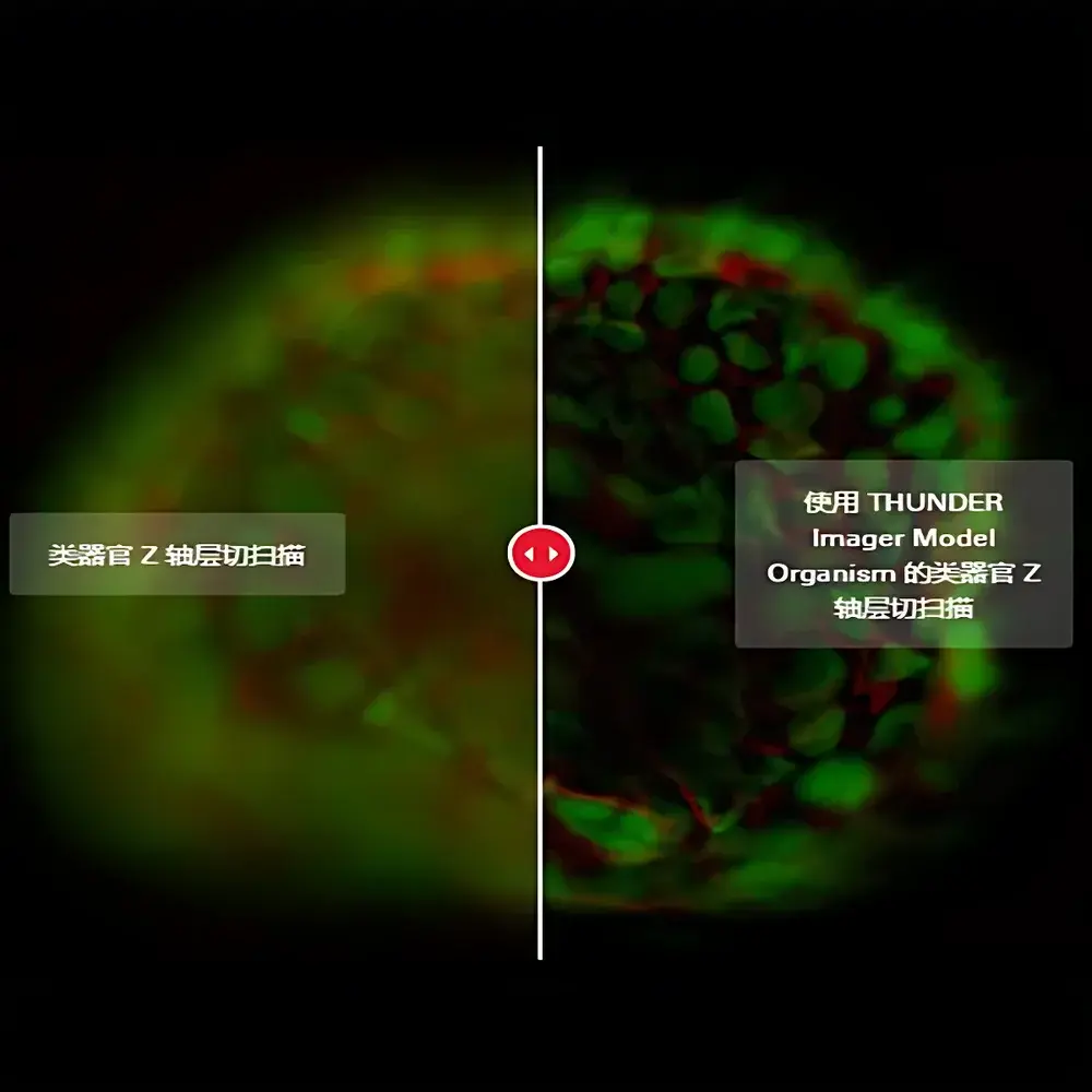
- Instant Computational Clearing (ICC): Real-time background suppression and depth-resolved contrast enhancement across full-field z-stacks—no post-acquisition processing latency.
- Hybrid Optical Design: Seamless integration of stereomicroscopy (for whole-organism navigation) and high-resolution epifluorescence (for subcellular detail), with motorized zoom, focus, and filter turrets.
- Automated Workflow Engine: Scriptable acquisition protocols for multi-position, multi-channel, multi-z time-lapse—fully compatible with Leica Application Suite X (LAS X) and third-party scheduling tools.
- Physiology-First Imaging Chamber: Optional environmental control module maintains stable temperature (20–40 °C ±0.2 °C), humidity (>95% RH), and gas composition for long-term live imaging of sensitive models.
- Modular Camera Options: 20 MP color CMOS for broad-spectrum phenotyping; scientific CMOS (sCMOS) with >82% QE and sub-electron read noise for low-light dynamics such as calcium signaling or mitochondrial trafficking.
- Optimized Optics Suite: Includes correction-collar 2× objective for aqueous immersion, FluoCombi III high-NA objectives (0.5–1.0 NA), and THUNDER-optimized emission filters with <5 nm bandwidths.
Sample Compatibility & Compliance
The THUNDER Imager Model Organism supports native, cleared, and embedded specimens up to 15 mm in height and 50 mm in lateral dimension—including whole zebrafish (up to 7 dpf), dissected murine organs (e.g., brain, kidney), hydrogel-embedded organoids (~100–300 µm), and ScaleS-cleared E12–E14 mouse embryos. It accommodates standard 35 mm dishes, 6–to-96-well plates, and custom chamber slides. All optical components comply with ISO 10934-1 (microscope terminology) and ISO/IEC 2382:2015 (information technology—vocabulary), ensuring traceable definitions of resolution, contrast, and signal fidelity. Data provenance meets GLP/GMP-aligned metadata standards: each image embeds EXIF tags for acquisition parameters (exposure, gain, z-step, objective ID), instrument serial number, and user-defined experimental annotations. Optional FDA 21 CFR Part 11 compliance packages include electronic signatures, audit trails, and role-based access control via LAS X Enterprise.
Software & Data Management
Leica LAS X 4.x software provides unified control of hardware, acquisition, and computational processing. The THUNDER 3D Workstation includes dedicated modules for ICC parameter optimization, batch z-stack reconstruction, intensity normalization across time points, and ROI-based quantification (e.g., fluorescence intensity, object count, morphometric profiling). Export formats include OME-TIFF (with full metadata), HDF5 (for large-scale time-series), and N5 (compatible with BigDataViewer and napari). Integration with Python (via PyLeica SDK) and MATLAB enables custom algorithm deployment—such as neural network–based segmentation or motion correction—within the native acquisition loop. Raw data storage follows FAIR principles (Findable, Accessible, Interoperable, Reusable), with optional linkage to institutional LIMS or ELN systems through RESTful API endpoints.
Applications
- Transgenic line characterization: Rapid phenotypic screening of reporter expression patterns across developmental stages in zebrafish or C. elegans.
- Neurodevelopmental dynamics: Long-term tracking of axon guidance, synapse formation, and microglial motility in intact larval brains under physiological conditions.
- Organoid maturation analysis: Quantitative assessment of lumen formation, cell polarity, and vascular network integration in human iPSC-derived gut or cerebral organoids.
- Plant morphogenesis: Time-resolved imaging of root gravitropism, stomatal development, or pathogen response in Arabidopsis thaliana seedlings without dissection.
- Clear-tissue validation: Correlative assessment of antibody penetration, labeling efficiency, and structural preservation in CLARITY- or CUBIC-processed murine tissues.
FAQ
How does Instant Computational Clearing differ from conventional deconvolution?
ICC performs iterative deconvolution during image acquisition—not after—and uses a calibrated, specimen-adapted PSF model. This avoids the information loss and artifact amplification common in post-hoc blind deconvolution.
Can the system image non-cleared, opaque specimens?
Yes. While optimal for cleared or semi-transparent samples, ICC significantly improves contrast in native zebrafish larvae or Drosophila pupae by suppressing scattered light—enabling usable depth penetration up to 200 µm.
Is environmental control integrated or add-on?
A modular environmental chamber is available as an optional accessory, fully synchronized with LAS X for closed-loop regulation and logging.
What file formats are supported for downstream analysis in Imaris or Arivis?
OME-TIFF export preserves all metadata and channel alignment; HDF5 files support direct loading into Imaris via the Bio-Formats importer and Arivis Vision4D via native HDF5 plugin.
Does the system support multi-user workflow management?
Yes. LAS X Enterprise supports project-level permissions, instrument queue scheduling, and version-controlled protocol libraries accessible across networked workstations.
Related Products


