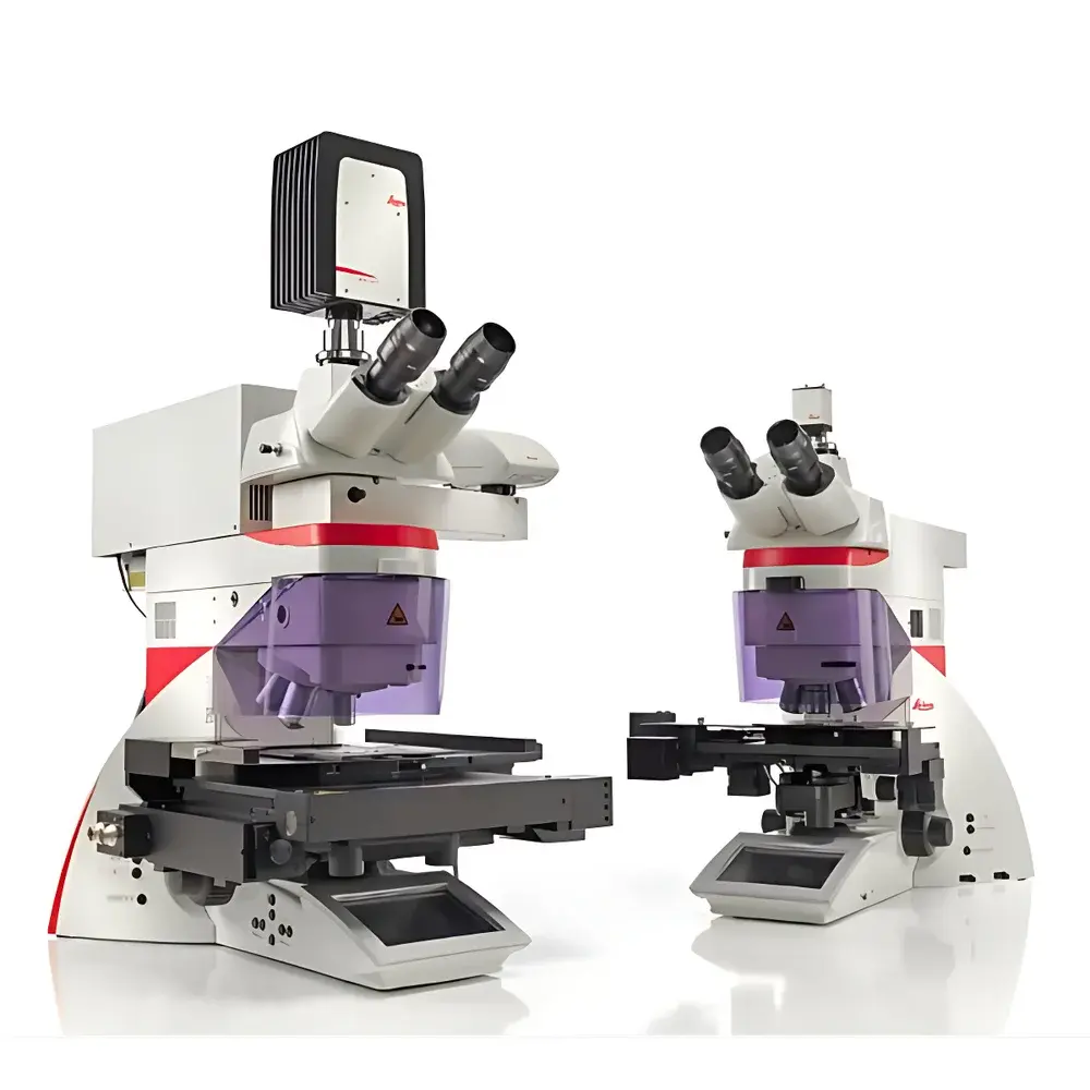
Leica LMD6 & LMD7 Laser Microdissection Systems
| Brand | Leica |
|---|---|
| Origin | Germany |
| Manufacturer Type | Authorized Distributor |
| Product Category | Imported Instrument |
| Model | Leica LMD6 & LMD7 Laser Microdissection Systems |
| Instrument Type | Precision Laser Microdissection System |
| Cutting Principle | Beam-Scanning (Non-Contact, Gravity-Based Collection) |
| Optical Architecture | Upright Microscope Platform with Galvo-Steered UV Laser (355 nm) |
| Software Platform | Leica Application Suite (LAS) X v7.6 with Workflow-Oriented Modules |
Overview
The Leica LMD6 and LMD7 Laser Microdissection (LMD) systems are high-precision, non-contact tissue isolation platforms engineered for spatially resolved molecular analysis. Unlike mechanical or manual microdissection methods, these systems employ a pulsed ultraviolet (UV) laser—typically at 355 nm—focused through an upright microscope optical path to ablate defined regions directly from heterogeneous tissue sections or cultured cells. Critically, the LMD6/LMD7 implement a beam-scanning paradigm: the laser spot is steered via high-speed galvanometric mirrors across the sample surface while the specimen remains stationary on the stage. This eliminates mechanical drift, stage vibration, and positional uncertainty inherent in sample-moving architectures—ensuring micron-level reproducibility and preserving morphological integrity of adjacent structures. The ablated material is collected solely by gravity into sterile, low-binding receptacles (e.g., PCR tube caps, 8-strip tubes, or specialized collection plates), eliminating cross-contamination risks associated with contact-based transfer or adhesive cap-based capture.
Key Features
- Beam-Scanning Architecture: Galvo-driven UV laser scanning enables precise, real-time contour tracing without stage movement—ideal for delicate or unstained samples requiring continuous visual monitoring.
- Gravity-Based Collection: No adhesives, no caps, no vacuum—ablated tissue fragments fall freely under gravity into designated collection vessels, ensuring 100% contamination-free downstream extraction for genomics, transcriptomics, proteomics, and metabolomics.
- Upright Microscope Platform: Compatible with standard glass slides, PEN-membrane slides, PET slides, and ibidi µ-Slides—supporting both fixed and live-cell applications including time-lapse imaging during dissection.
- Integrated Environmental Control: Optional climate chamber integration maintains physiological conditions (temperature, CO₂, humidity) for live-cell LMD workflows, enabling single-cell cloning, re-culturing, or functional assays post-isolation.
- Optimized Slide Compatibility: PET slides minimize plasticizer leaching for proteomic/metabolomic workflows; DIRECTOR slides enable film-free cutting for ultra-low background analyses; PEN membranes support RNA integrity preservation.
Sample Compatibility & Compliance
The LMD6 and LMD7 accommodate a broad spectrum of sample formats: cryosections (4–20 µm), FFPE sections (1–10 µm), cytospins, cell smears, and adherent monolayers grown on coated or uncoated surfaces. All workflows comply with Good Laboratory Practice (GLP) documentation requirements. The LAS X software supports audit trails, user access control, and electronic signature functionality aligned with FDA 21 CFR Part 11 guidelines when configured with appropriate IT infrastructure. Data export conforms to MIAME-compliant metadata standards, facilitating integration into LIMS environments and bioinformatics pipelines. ISO 13485-certified manufacturing ensures traceability of optical components and laser calibration records.
Software & Data Management
Leica Application Suite (LAS) X v7.6 provides a workflow-centric interface designed specifically for microdissection tasks. Users navigate via intuitive touchscreen or mouse input to define regions of interest (ROIs) using polygon, freehand, or auto-detection tools—including Adaptive Vision Classification (AVC) for unsupervised cell-type segmentation based on texture and intensity features. Real-time laser control, motorized focus tracking, and synchronized time-lapse recording (up to 4K resolution) are fully integrated. Project metadata—including laser parameters (pulse energy, frequency, dwell time), ROI coordinates, and collection vessel mapping—are embedded in exported TIFF/OME-TIFF files. Optional modules include database linking for sample annotation, batch processing scripts for high-throughput studies, and DICOM export for pathology lab interoperability.
Applications
- Single-Cell & Subcellular Genomics: Isolation of nuclei, mitotic figures, or specific epithelial compartments from tumor heterogeneity studies.
- Transcriptomics of Rare Cell Populations: Retrieval of in situ mRNA-expressing neurons, immune infiltrates, or stromal subtypes without amplification bias.
- Proteomic Biomarker Discovery: Film-free DIRECTOR slide cutting minimizes keratin and polymer interference in LC-MS/MS workflows.
- Live-Cell Functional Analysis: Targeted ablation of individual cells within co-cultures followed by re-plating and phenotypic readout.
- Forensic & Archaeological Tissue Analysis: Contamination-free retrieval of degraded DNA from mixed or aged specimens.
FAQ
How does beam-scanning differ from cap-based LMD systems?
Beam-scanning eliminates physical contact between laser and collection surface—removing risks of cap contamination, thermal degradation, or incomplete transfer. Gravity collection preserves native biomolecule integrity.
Can the LMD6/LMD7 be used for live-cell dissection under controlled environmental conditions?
Yes—when coupled with an optional incubation chamber, the system supports CO₂-regulated, temperature-stabilized dissection of adherent or suspension cultures with real-time phase contrast or fluorescence monitoring.
Which slide types are recommended for proteomics applications?
PET-coated slides (low plasticizer content) or DIRECTOR slides (no polymer film) are validated for minimal background interference in mass spectrometry workflows.
Is LAS X software compliant with regulatory data integrity requirements?
When deployed with role-based permissions, electronic signatures, and audit trail logging enabled, LAS X meets core elements of FDA 21 CFR Part 11 and EU Annex 11 for analytical instrument software.
What is the typical lateral resolution achievable with the UV laser?
The diffraction-limited spot size at 355 nm yields nominal lateral resolution of ≤1 µm, with practical cutting line widths adjustable between 0.8 µm and 5 µm depending on tissue type and laser fluence settings.


