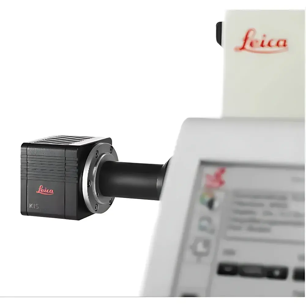
Leica K5 sCMOS Microscope Camera
| Brand | Leica |
|---|---|
| Origin | Germany |
| Model | K5 |
| Sensor Type | Scientific CMOS (sCMOS) |
| Resolution | 2048 × 2048 (4.2 MP) |
| Maximum Frame Rate | 40 fps (full resolution) |
| Quantum Efficiency | up to 80% @ 550 nm |
| Pixel Size | 6.5 µm |
| Read Noise | <1.1 e⁻ (typ.) |
| Dynamic Range | >30,000:1 |
| Interface | Camera Link HS or USB 3.2 Gen 2 (configurable) |
| Compliance | CE, RoHS, ISO 9001 certified manufacturing |
| Software Compatibility | Leica LAS X, THUNDER Imager platform, and third-party APIs (e.g., Micro-Manager, Python via SDK) |
Overview
The Leica K5 sCMOS Microscope Camera is a high-performance, scientific-grade imaging sensor engineered for demanding fluorescence microscopy applications in life science research laboratories. Built upon a back-illuminated sCMOS architecture, the K5 delivers exceptional photon detection efficiency, low read noise, and high-speed acquisition—enabling quantitative widefield imaging with superior signal fidelity. Its core measurement principle relies on pixel-level charge integration under controlled exposure, followed by correlated double sampling and high-fidelity analog-to-digital conversion—optimized to preserve spatial and temporal integrity across dynamic biological processes. Designed as a native imaging engine for the Leica THUNDER Imager platform, the K5 supports computational clearing workflows that suppress out-of-focus blur in real time without requiring hardware-based optical sectioning. This makes it particularly suitable for live-cell imaging, 3D organoid analysis, multi-channel immunofluorescence, and time-lapse studies where phototoxicity minimization and temporal resolution are critical.
Key Features
- Back-illuminated sCMOS sensor with 4.2 megapixel resolution (2048 × 2048), delivering high spatial fidelity for subcellular structure visualization.
- Quantum efficiency exceeding 80% at 550 nm—maximizing photon capture from common fluorophores such as Alexa Fluor 488, DAPI, MitoTracker Red, and Alexa Fluor 647.
- Read noise below 1.1 electrons (typical), enabling reliable detection of low-intensity signals in dimly labeled specimens or sparse expression systems.
- Full-frame acquisition at up to 40 frames per second—facilitating high-throughput time-lapse imaging and rapid event capture in motile cells or calcium transients.
- Dynamic range greater than 30,000:1—supporting simultaneous visualization of both bright and dim structures within the same field of view without saturation or loss of detail.
- Configurable interface options: Camera Link HS for ultra-low latency and deterministic timing in synchronized multi-modal setups, or USB 3.2 Gen 2 for simplified integration into standard workstation environments.
- Firmware-upgradable architecture with embedded calibration tables for gain, offset, and flat-field correction—ensuring long-term measurement consistency across instruments and users.
Sample Compatibility & Compliance
The Leica K5 is compatible with all Leica DM series upright and inverted research microscopes equipped with C-mount or F-mount adapters, and integrates seamlessly with third-party microscope platforms via standardized mechanical and electrical interfaces. It supports multi-channel fluorescence imaging across UV–NIR spectral bands (350–1000 nm), including DAPI, FITC, TRITC, Cy5, and far-red dyes. The camera complies with CE marking requirements for electromagnetic compatibility and safety (EN 61326-1, EN 61000-6-3), RoHS Directive 2011/65/EU, and is manufactured under an ISO 9001-certified quality management system. When used within validated THUNDER workflows, image acquisition metadata—including exposure time, gain, binning, and timestamp—is automatically embedded in TIFF and OME-TIFF files, supporting GLP/GMP-aligned documentation practices and audit readiness per FDA 21 CFR Part 11 when paired with compliant LIMS or ELN systems.
Software & Data Management
The K5 operates natively within Leica LAS X software and the THUNDER Imager platform, providing full control over exposure parameters, region-of-interest selection, hardware-triggered acquisition, and real-time computational clearing. It also supports open-standard APIs—including Micro-Manager device adapter and Python-based SDK—for custom script development, automated assay pipelines, and integration into AI-driven image analysis frameworks. All acquired images include embedded EXIF and OME metadata, ensuring traceability of acquisition conditions. Time-series datasets can be exported in standardized formats (OME-TIFF, HDF5) compatible with Fiji/ImageJ, Icy, QuPath, and commercial AI training platforms. Audit trails for parameter changes and acquisition logs are retained locally or configurable for network storage, aligning with laboratory data integrity principles.
Applications
- Live-cell fluorescence imaging of cytoskeletal dynamics, mitochondrial trafficking, and nuclear translocation events.
- High-content screening of 3D cell cultures and organoids using multi-color immunolabeling protocols.
- Time-lapse quantification of proliferation, migration, and apoptosis in co-culture models.
- Widefield deconvolution-enhanced imaging for routine histopathology and developmental biology studies.
- Correlative light microscopy (CLM) workflows where precise synchronization with electrophysiology or microfluidic stimulation is required.
- Training and validation of deep learning models for segmentation and classification—leveraging its high SNR and reproducible pixel response.
FAQ
Is the Leica K5 compatible with non-Leica microscope systems?
Yes—the K5 supports standard C-mount mechanical coupling and offers programmable GPIO triggers and TTL synchronization, enabling integration with Nikon, Olympus, Zeiss, and custom-built optical platforms via third-party drivers or SDK-based development.
Does the K5 support binning or region-of-interest (ROI) readout?
Yes—hardware binning (2×2, 4×4) and arbitrary ROI selection are supported, allowing trade-offs between speed, sensitivity, and field-of-view depending on experimental needs.
How is calibration maintained across long-term use?
The K5 includes factory-characterized flat-field and dark-current maps stored in onboard memory; these are applied automatically during acquisition. Users may perform optional recalibration using Leica’s provided uniform illumination source and LAS X calibration module.
Can the K5 be used for quantitative intensity measurements?
Yes—its linear response over six orders of magnitude, combined with calibrated EMVA 1288-compliant performance metrics (gain, quantum efficiency, noise), supports absolute intensity quantification when used with standardized reference samples and controlled illumination.
What support is available for developers integrating the K5 into custom software?
Leica provides a comprehensive C++ and Python SDK, detailed API documentation, example code repositories, and technical support through its Scientific Imaging Developer Program.


