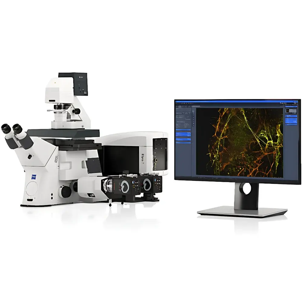
ZEISS Elyra 7 Cross-Scale Super-Resolution Microscope
| Brand | ZEISS |
|---|---|
| Origin | Germany |
| Model | Elyra 7 |
| Category | Imported Instrument |
| Distribution Type | Authorized Distributor |
| Pricing | Available Upon Request |
Overview
The ZEISS Elyra 7 is a modular, cross-scale super-resolution microscope engineered for quantitative multimodal imaging across biological length scales—from intact tissue sections down to single-molecule localization. It integrates structured illumination microscopy (SIM), total internal reflection fluorescence (TIRF), and single-molecule localization microscopy (SMLM) within a single optical platform, enabling seamless transitions between widefield, optical sectioning, and nanoscale resolution modes. Its core architecture leverages ZEISS’s Lattice SIM technology, which employs a high-contrast, low-phototoxicity excitation pattern to achieve lateral resolution of ≤60 nm in live or fixed specimens—without requiring specialized fluorophores or extreme labeling densities. Unlike conventional confocal or STED systems, the Elyra 7 maintains compatibility with standard organic dyes (e.g., Alexa Fluor, CF dyes) and genetically encoded tags (e.g., mEos, Dronpa), supporting longitudinal studies under physiological conditions. The system is designed for reproducible, correlative workflows where spatial registration between macroscopic tissue context and sub-diffraction molecular architecture is essential—for example, mapping synaptic protein distributions relative to dendritic morphology in brain slices, or correlating vascular network topology with pericyte positioning in intestinal epithelium.
Key Features
- Lattice SIM mode delivering ≤60 nm lateral resolution in live-cell imaging with minimal photobleaching and enhanced optical sectioning capability
- Integrated TIRF illumination for evanescent-field imaging of membrane-proximal events (e.g., vesicle docking, receptor clustering)
- Full SMLM acquisition and reconstruction pipeline—including dSTORM, PALM, and multi-color single-molecule tracking—with real-time drift correction and sub-pixel localization precision
- Motorized, software-synchronized objective turret supporting simultaneous use of low-magnification (10×/0.3 NA) objectives for overview imaging and high-NA oil immersion (63×/1.4 NA) objectives for nanoscale detail
- Multi-channel spectral unmixing and precise chromatic alignment calibration across ≥4 fluorescence channels, validated using NIST-traceable reference beads
- Modular design compliant with ISO 13322-2 (particle size analysis by optical microscopy) and ASTM E2859 (standard guide for fluorescence microscopy image acquisition)
Sample Compatibility & Compliance
The Elyra 7 supports a broad range of specimen formats: thick (>100 µm) cleared tissues, adherent cultured cells, suspension cells on functionalized coverslips, and ultrathin cryosections. It accommodates standard #1.5H glass coverslips (0.17 mm thickness) and is compatible with common mounting media (e.g., ProLong Diamond, Vectashield). All imaging modalities comply with GLP/GMP-relevant documentation requirements: audit trails for acquisition parameters (exposure time, laser power, gain settings), user authentication logs, and timestamped metadata export in OME-TIFF format. The system meets IEC 61000-6-3 (EMC emissions) and IEC 61000-6-2 (immunity) standards for laboratory instrumentation. For regulated environments, optional 21 CFR Part 11-compliant software modules provide electronic signatures, role-based access control, and immutable data archiving.
Software & Data Management
ZEISS ZEN Imaging Software (Blue edition) serves as the unified control and analysis environment. It enables synchronized hardware triggering, real-time SMLM localization rendering, and automated multi-scale stitching (e.g., merging 10× whole-tissue scans with 63× SMLM ROIs). Data management follows FAIR principles: raw datasets retain full metadata (including objective serial numbers, laser calibration timestamps, and environmental sensor logs), and processed results are exportable in HDF5 or OME-Zarr formats for integration into institutional LIMS or cloud-based analysis pipelines (e.g., Napari, QuPath, BigDataViewer). Batch processing scripts support standardized quantification of cluster density, nearest-neighbor distances, and co-localization coefficients (Manders’ M1/M2, Pearson’s r) across hundreds of fields of view.
Applications
- Correlative tissue-to-molecule imaging: e.g., mapping SYCP3/SYCP1 nanoscale spacing (<60 nm) within meiotic chromosome axes in murine testis sections
- Dynamic subcellular trafficking: dual-color 2D STORM of microtubules and TOMM20 in Cos-7 cells to resolve mitochondrial docking sites relative to cytoskeletal anchors
- Vascular–neural interface mapping: simultaneous Alexa 488 (vasculature) and Alexa 647 (neuronal processes) labeling in murine small intestine, registered across 10× and 63× magnifications
- Drug-induced ultrastructural remodeling: time-lapse Lattice SIM monitoring of nuclear pore complex reorganization during kinase inhibitor treatment
- Quantitative multiplexed biomarker validation: SMLM-based stoichiometry analysis of receptor dimerization states in primary human tumor biopsies
FAQ
Does the Elyra 7 require special fluorophores for SMLM operation?
No—standard organic dyes (e.g., Alexa Fluor 647, CF568) and photoactivatable proteins (e.g., mEos3.2) are fully supported. Buffer optimization (e.g., oxygen scavenging systems) is recommended but not mandatory.
Can I perform live-cell SMLM with the Elyra 7?
Yes—using low-intensity activation lasers and fast EMCCD/sCMOS readout, sub-second temporal resolution is achievable for selected targets such as clathrin-coated pit dynamics.
How is multi-scale image registration performed?
Hardware-based stage encoding and software-driven fiducial marker detection (e.g., fluorescent beads embedded in mounting medium) enable <100 nm positional accuracy across magnifications.
Is the system compatible with third-party analysis tools?
Yes—raw data exports include complete metadata in OME-TIFF/HDF5; plugins exist for ImageJ/Fiji, MATLAB, and Python (via zarr, tifffile, and ome-zarr libraries).
What service and compliance documentation is provided?
Each system ships with Factory Acceptance Test (FAT) report, ISO 17025-accredited calibration certificate for objective point spread function (PSF), and CE/UKCA declarations of conformity.


