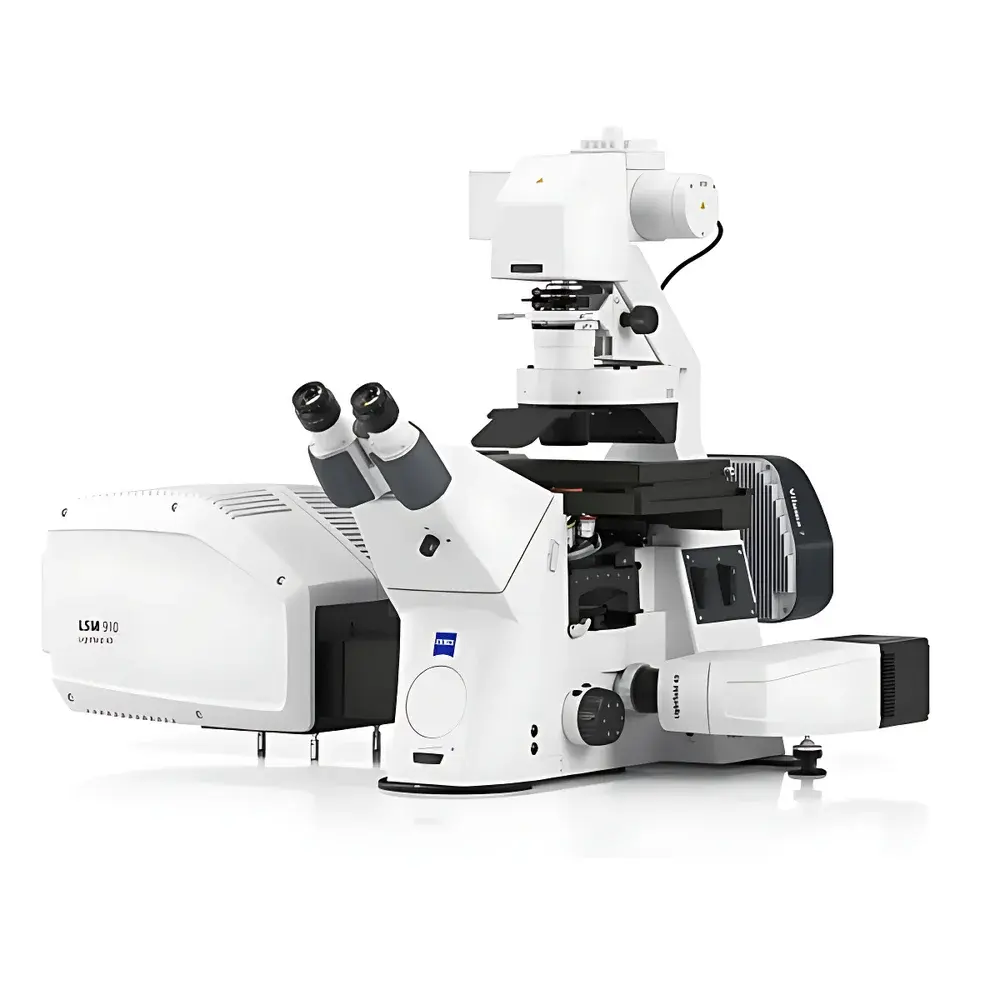
ZEISS LSM 910 Laser Scanning Confocal Microscope
| Brand | ZEISS |
|---|---|
| Origin | Germany |
| Model | LSM 910 |
| Instrument Type | Point-Scanning Confocal Microscope |
| Optical Resolution (XY) | ≤ 80 nm (measured via FWHM of fluorescent beads) |
| Volumetric Imaging Speed | Up to 80 volumes/s |
| System Architecture | Integrated Airyscan Detection |
| Compliance | Designed for GLP/GMP-aligned workflows, compatible with FDA 21 CFR Part 11–enabled software configurations |
| Software Platform | ZEN Intellesis & ZEN Blue with LightField 4D module |
Overview
The ZEISS LSM 910 Laser Scanning Confocal Microscope is a high-performance point-scanning confocal system engineered for quantitative, multi-dimensional fluorescence imaging in life science laboratories. Based on the fundamental principle of optical sectioning—where a focused laser beam scans the specimen point-by-point while a conjugate pinhole rejects out-of-focus light—the LSM 910 delivers intrinsic axial resolution and high signal-to-noise ratio (SNR) without computational deconvolution. Its core optical architecture integrates a modular galvo-resonant scanning unit, high-efficiency spectral detection pathways, and an Airyscan-enabled detector array optimized for photon-limited conditions. The system supports both conventional confocal mode and Airyscan super-resolution mode (with lateral resolution down to ≤80 nm, validated using calibrated fluorescent nanospheres), enabling rigorous morphological and spatial quantification across fixed and live biological specimens.
Key Features
- Point-scanning confocal architecture with dual-galvo and resonant scanning options for flexible speed/resolution trade-offs
- Airyscan detection system delivering up to 1.7× improvement in signal intensity and ≥1.7× enhancement in lateral resolution compared to standard confocal detection
- Simultaneous multi-channel spectral unmixing across 32 channels (340–950 nm range) with tunable bandpass selection
- LightField 4D acquisition engine supporting real-time volumetric imaging at up to 80 volumes per second—enabling dynamic tracking of subcellular processes in live embryos and organoids
- Integrated environmental control interface for CO₂, temperature, and humidity regulation—compatible with long-term live-cell imaging chambers
- Compact footprint (W × D × H: 720 × 650 × 630 mm) minimizing benchtop space requirements without compromising optical path stability or thermal management
- ZEN Blue and ZEN Intellesis software suite with AI-assisted segmentation, batch processing pipelines, and reproducible acquisition parameter templating
Sample Compatibility & Compliance
The LSM 910 accommodates a broad range of biological specimens—from single mammalian cells and primary neuronal cultures to thick tissue sections (up to 200 µm), cleared whole-mount organs, and 3D organoid models. It supports immersion objectives (oil, glycerol, water) with correction collars for depth-dependent spherical aberration compensation. All hardware and firmware are designed to meet CE marking requirements under Directive 2014/30/EU (EMC) and 2014/35/EU (LVD). When deployed with ZEN Blue v3.7+ and configured audit-trail logging, the system supports compliance with FDA 21 CFR Part 11 for electronic records and signatures. Routine calibration procedures align with ISO/IEC 17025–recommended traceability frameworks for fluorescence intensity and spatial calibration standards.
Software & Data Management
Acquisition, analysis, and data archival are unified within the ZEN software ecosystem. ZEN Blue provides intuitive wizard-driven experiment setup, including automated focus maintenance (Definite Focus 2), adaptive illumination control (Smart Setup), and hardware-synchronized time-lapse registration. ZEN Intellesis introduces deep learning–based object detection and classification trained on community-validated ground-truth datasets (e.g., BBBC, CellTracking Challenge). Raw data are stored in OME-TIFF format compliant with the Open Microscopy Environment (OME) metadata schema, ensuring interoperability with ImageJ/Fiji, QuPath, and Python-based analysis pipelines (e.g., napari, scikit-image). Project-level metadata—including objective ID, laser power history, pinhole position, and environmental sensor logs—is embedded and exportable for GLP audit readiness.
Applications
The LSM 910 serves as a foundational platform for hypothesis-driven research in cell biology (e.g., mitotic spindle dynamics, organelle contact site mapping), developmental biology (e.g., cardiac morphogenesis in zebrafish embryos), neurobiology (e.g., dendritic spine turnover, synaptic vesicle trafficking), and translational oncology (e.g., tumor spheroid invasion assays). Its four-dimensional imaging capability enables quantitative hemodynamic profiling in vascularized organoids; its molecular dynamics tools support diffusion coefficient estimation (via FRAP, FCS, or single-particle tracking) and binding kinetics modeling in native cellular environments. Cross-disciplinary applications include biophysical characterization of hydrogel–cell interfaces and structural analysis of plant vascular bundles under stress conditions.
FAQ
What is the maximum achievable lateral resolution in Airyscan mode?
Measured using 100-nm fluorescent beads and full-width-at-half-maximum (FWHM) analysis, the system achieves ≤80 nm lateral resolution under optimal labeling and immersion conditions.
Can the LSM 910 perform simultaneous multi-color imaging with spectral unmixing?
Yes—it supports real-time spectral detection across 32 channels with linear unmixing of fluorophores exhibiting ≥25 nm spectral separation (e.g., GFP/mCherry/CFP combinations).
Is the system compatible with third-party incubation systems?
It features standardized electrical and pneumatic interfaces (including RS-232, TTL triggers, and analog voltage outputs) for seamless integration with commercial stage-top and chamber-based live-cell systems.
Does ZEN software support automated batch analysis of time-lapse datasets?
Yes—ZEN Intellesis includes scriptable macro workflows for ROI-based intensity quantification, motion-corrected stack alignment, and morphology parameter extraction across hundreds of time points.
How is phototoxicity minimized during long-term live imaging?
Through hardware-level laser power modulation, adaptive dwell time adjustment, and low-noise GaAsP detectors that maximize photon collection efficiency—reducing required excitation intensity by up to 50% versus conventional PMT-based systems.


