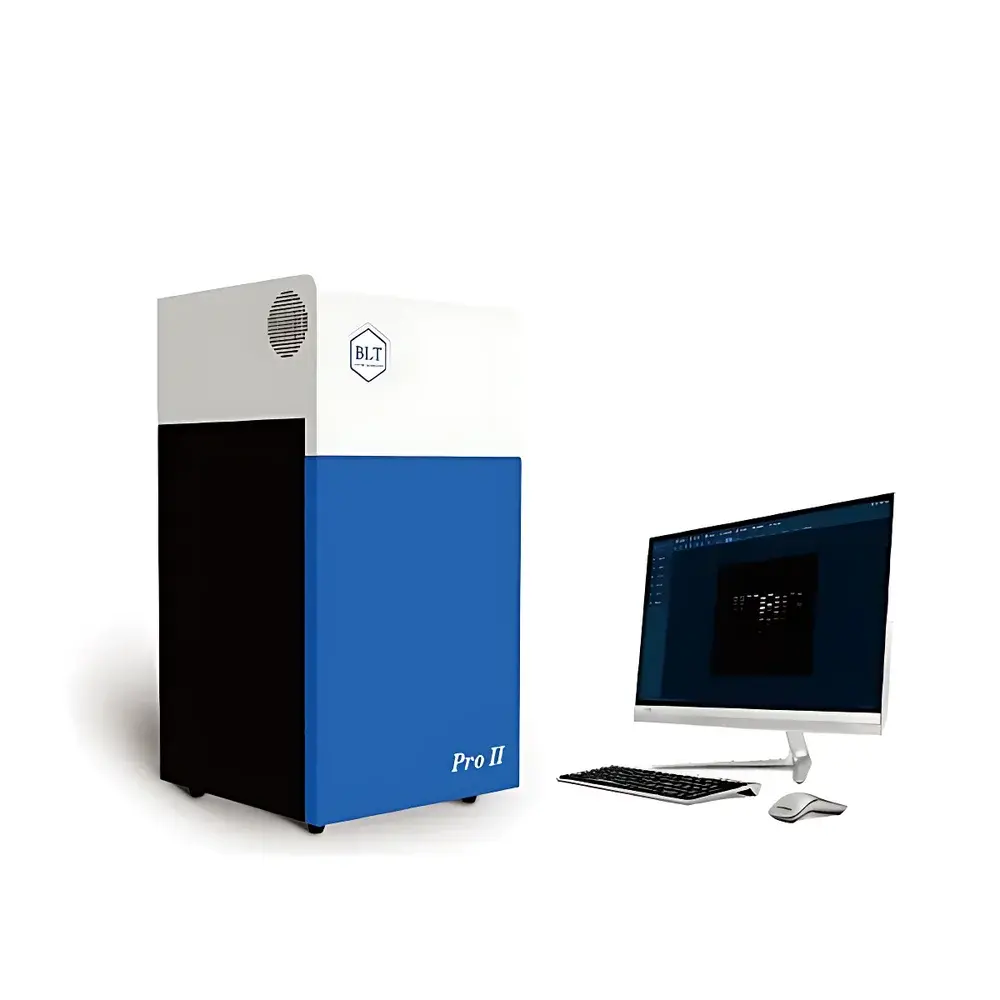
BioLight GelView 1500ProⅡ Fully Automated Gel Imaging System
| Brand | BioLight |
|---|---|
| Origin | Guangdong, China |
| Manufacturer Type | Authorized Distributor |
| Instrument Category | Domestic |
| Model | GelView 1500ProⅡ |
| Imaging Mode | Chemiluminescence & Multi-Excitation Gel Imaging |
| CCD Resolution | 1.4 Megapixels (Effective) |
| Lens | F1.2, 8–48 mm Motorized Zoom with Auto-Focus |
| UV Safety Features | Door-Interlocked UV Shut-off & Programmable UV Delay Timer |
| Illumination Options | UV Transillumination (302 nm), Blue Light Transillumination (470 nm), White Light Reflective Illumination |
| Sample Compatibility | Agarose Gels, SDS-PAGE Gels, Petri Dishes, 96-Well Plates, Colony Arrays, Antibiotic Zone Assays |
| Software Compliance | GLP-Compliant Multi-User Audit Trail & Data Integrity Controls |
Overview
The BioLight GelView 1500ProⅡ Fully Automated Gel Imaging System is a high-integrity, multi-modal imaging platform engineered for quantitative analysis of nucleic acid and protein electrophoresis gels under ultraviolet (UV), blue-light, and visible-light excitation conditions. Based on solid-state CCD detection and precision optical path design, the system employs a scientific-grade 1.4-megapixel monochrome CCD sensor with high quantum efficiency in the 300–600 nm range, enabling low-noise acquisition of both ethidium bromide (EB)-stained DNA and SYBR® Green-labeled amplicons, as well as Coomassie Brilliant Blue- or silver-stained protein gels. Its modular illumination architecture integrates switchable UV transilluminators (302 nm), high-output blue-light transilluminators (470 nm), and uniform white LED reflective lighting—each independently controlled and interlocked with mechanical door sensors to ensure operator safety and regulatory compliance. The system operates without manual focus adjustment, relying instead on a motorized F1.2 wide-aperture zoom lens (8–48 mm) with closed-loop auto-focus calibration prior to each exposure.
Key Features
- Fully automated imaging workflow: One-touch acquisition, auto-exposure optimization, and real-time preview reduce user variability and training burden.
- Regulatory-ready hardware safety: UV lamp automatically deactivates upon door opening; programmable UV delay timer prevents unnecessary exposure during sample loading.
- Laser-guided positioning system: Dual crosshair lasers project onto the imaging surface to ensure precise centering of gels, Petri dishes, or microplates within the field of view—critical for reproducible ROI definition across multiple users and sessions.
- Modular illumination tray design: UV and blue-light transilluminators are mechanically interchangeable without tools, enabling rapid reconfiguration between nucleic acid and protein imaging protocols.
- High-dynamic-range acquisition: 16-bit analog-to-digital conversion supports linear quantification over 4 orders of magnitude intensity range—essential for comparative band densitometry and semi-quantitative Western blot analysis.
- Integrated thermal management: Active cooling maintains CCD junction temperature ≤ –25°C below ambient, minimizing dark current noise during long-exposure chemiluminescent imaging.
Sample Compatibility & Compliance
The GelView 1500ProⅡ accommodates standard electrophoretic formats including 10 × 10 cm to 20 × 20 cm agarose and polyacrylamide gels, as well as non-gel substrates such as bacterial colony arrays on Petri dishes, antibiotic diffusion zone assays on Mueller-Hinton agar, and 96-well microtiter plates used in ELISA or cell viability screening. It supports all major fluorescent and chromogenic stains—including EB, GoldView™, GeneFinder™, SYBR® Safe, SYBR® Gold, Coomassie R-250, Deep Purple™, and silver nitrate—with optimized exposure presets stored per stain type. The system complies with IEC 61010-1 for laboratory equipment safety and incorporates mechanical and firmware-level UV interlocks meeting EN 60825-1 requirements. Its software architecture supports ALCOA+ data integrity principles and includes full audit trail logging (user ID, timestamp, parameter changes, image annotations) required for GLP and ISO/IEC 17025 environments.
Software & Data Management
BioLight GelView Analysis Suite v5.x provides a validated, role-based interface with configurable user permissions (administrator, technician, reviewer). All image acquisition parameters—including exposure time, binning mode, lens zoom position, and illumination source—are embedded in TIFF metadata and cannot be altered post-acquisition. Quantitative modules include lane/band detection with background subtraction algorithms compliant with NIH ImageJ methodology benchmarks, colony counting using adaptive threshold segmentation, zone-of-inhibition measurement per CLSI M02-A13 guidelines, and microplate well intensity profiling with coefficient-of-variation reporting. Raw and processed data export options include CSV, PDF reports with embedded digital signatures, and DICOM-compatible encapsulation for LIMS integration. Software validation documentation (IQ/OQ/PQ protocols) and 21 CFR Part 11-compliant electronic signature modules are available upon request.
Applications
- Quantitative analysis of PCR products, restriction digests, and plasmid preparations on agarose gels.
- Densitometric evaluation of SDS-PAGE-separated proteins, including phosphorylation state assessment via Pro-Q Diamond staining.
- Chemiluminescent detection of horseradish peroxidase (HRP)-conjugated antibodies in Western blots with sub-femtogram sensitivity.
- High-throughput colony enumeration and morphological classification in microbial strain isolation workflows.
- Antibiotic potency testing via disk diffusion assays (Kirby-Bauer method) with automatic zone diameter measurement and CLSI breakpoint interpretation.
- Dot/slot blot hybridization signal quantification and normalization against housekeeping gene controls.
FAQ
Does the GelView 1500ProⅡ support fluorescence imaging with SYBR® Gold and SYBR® Safe?
Yes—the system’s 470 nm blue-light transilluminator is spectrally matched to the excitation maxima of both dyes, and its high-sensitivity CCD enables detection at sub-nanogram DNA levels.
Is the software validated for use in regulated pharmaceutical laboratories?
The GelView Analysis Suite supports 21 CFR Part 11 compliance when deployed with optional electronic signature and audit trail modules; IQ/OQ documentation packages are provided for GxP environments.
Can the system acquire images from X-ray film or autoradiography cassettes?
No—it is not configured for phosphorimaging or radioisotope detection; it is optimized for optical fluorescence, chemiluminescence, and reflectance imaging only.
What is the maximum gel size supported without cropping?
The active imaging area accommodates gels up to 20 × 20 cm; larger formats require stitching via the software’s multi-frame mosaic acquisition mode.
How is calibration maintained across multiple users and shifts?
Auto-calibration routines run at startup and before each acquisition sequence, verifying lens focus, illumination uniformity, and CCD gain offset—results are logged in the audit trail.


