CoreScanner Core Density X-ray Imaging & Elemental Analysis System
| Origin | Sweden |
|---|---|
| Manufacturer Type | Authorized Distributor |
| Origin Category | Imported |
| Model | CoreScanner |
| Pricing | Upon Request |
Overview
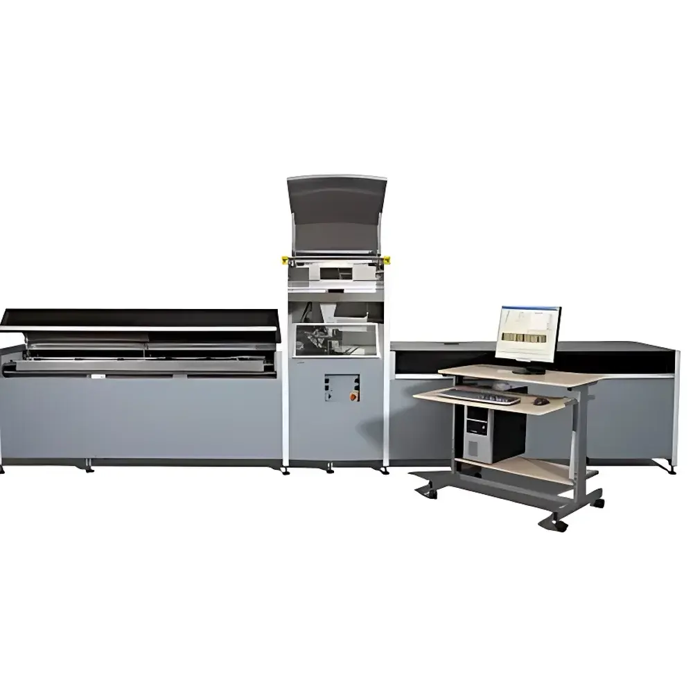
The CoreScanner Core Density X-ray Imaging & Elemental Analysis System is an industrial-grade, non-destructive micro-analytical platform engineered for high-resolution, multi-modal characterization of geological and environmental core samples. It integrates three complementary physical measurement principles: energy-dispersive X-ray fluorescence (ED-XRF) spectroscopy for quantitative elemental composition; digital X-ray micro-radiography for density and structural imaging; and high-fidelity orthogonal-polarized optical imaging for surface morphology and colorimetric texture analysis. Designed specifically for sedimentology, paleoclimatology, marine geoscience, and environmental forensics, the system enables centimeter- to micrometer-scale spatial profiling of intact cores—including split sediment cores, rock cores, peat monoliths, speleothems, and soil columns—without physical sectioning or chemical digestion. Its operational architecture complies with international radiation safety standards (IEC 61000-4, IEC 62471), and all X-ray subsystems are interlocked with real-time beam inhibition upon access door opening.
Key Features
- Triple-modality acquisition synchronized in a single pass: ED-XRF elemental mapping, X-ray attenuation-based density tomography, and RGB optical reflectance imaging
- High-sensitivity SDD (Silicon Drift Detector) cooled to –20 °C, enabling detection of elements from Al (13) to U (92) with <140 eV FWHM resolution at 5.9 keV (Mn-Kα)
- Dual-beam X-ray optics: collimated micro-focus beam (0.2 × 20 mm flat-field) for density radiography and a dedicated 0.1–0.2 mm spot XRF excitation beam
- Automated 1800 mm motorized sample stage with 20 µm positional repeatability and thermal drift compensation
- U-shaped sample cradle with five precisely indexed lateral positions (center ±10 mm, ±20 mm) for optimal beam alignment across heterogeneous core cross-sections
- Orthogonal-polarized 16-bit RGB CCD camera delivering 50 µm optical resolution in high-fidelity mode and 200 µm in rapid-scan mode over 100 mm field width
- Integrated radiation shielding compliant with EU Directive 2013/59/Euratom and equipped with fail-safe beam termination circuitry
- Robust thermal management via closed-loop water cooling system, supporting continuous operation >6000 hours/year
Sample Compatibility & Compliance
The CoreScanner accommodates a broad range of natural and prepared core geometries: split sediment cores up to 1750 mm in length and 120 mm in width; intact cylindrical specimens (e.g., stalagmites, tree cores, ice cores) with diameters ≤120 mm and thicknesses from 1–60 mm; and planar slabs such as polished thin sections or soil monoliths. Sample handling adheres to GLP-compliant workflows, and data provenance meets FDA 21 CFR Part 11 requirements for audit trail, electronic signature, and data integrity. The system supports traceable calibration using NIST-traceable reference materials (e.g., NIST SRM 2711a, USGS AGV-2), and analytical outputs are compatible with ISO/IEC 17025-accredited laboratory reporting frameworks. All XRF quantification routines incorporate matrix correction algorithms validated against ICP-MS and LOI-550 reference methods per ASTM D7348 and ISO 11885.
Software & Data Management
CoreScanner operation and data processing are managed through a suite of purpose-built, modular applications deployed on dual Windows-based workstations: Core Scanner Navigator for hardware orchestration and scan parameter definition; Qspec for spectral deconvolution, peak integration, and element-specific concentration modeling (including Compton/Rayleigh scattering ratio derivation for organic matter estimation); and ReDiCore for co-registered visualization of XRF, radiographic, and optical datasets in 2D line-scan or pseudo-3D volume reconstruction formats. Raw data files adhere to HDF5 format with embedded metadata (sample ID, timestamp, instrument configuration, calibration history), ensuring FAIR (Findable, Accessible, Interoperable, Reusable) compliance. Export options include ASCII, CSV, GeoTIFF, and NetCDF for downstream statistical analysis in R, Python (pandas/xarray), or MATLAB environments. Audit logs record every user action, parameter change, and calibration event with time-stamped, immutable entries.
Applications
- Paleoenvironmental reconstruction via high-resolution elemental stratigraphy (e.g., Fe/Ca/K/Si ratios as proxies for redox conditions, terrigenous input, biogenic productivity)
- Marine sediment provenance studies using rare earth element (REE) patterns and transition metal geochemistry
- Contaminant tracing in estuarine and lacustrine sediments (e.g., Pb, Zn, As, Hg accumulation profiles linked to industrial chronologies)
- Organic matter quantification through Compton-to-Rayleigh scattering intensity ratios (inc/coh), validated against LOI-550 and TOC measurements
- Volcanic tephra layer identification and correlation using Ti/Zr/Y signatures and glass shard morphology from optical imaging
- Soil carbon stock assessment and pedogenic horizon delineation in climate resilience research
- Non-invasive wood density profiling for dendrochronological and silvicultural studies
FAQ
What elements can the CoreScanner detect, and what is the typical detection limit?
The system detects elements from aluminum (Al) to uranium (U) using ED-XRF. Detection limits range from sub-ppm to low-ppm levels depending on element atomic number, matrix absorption, and acquisition time—validated against certified reference materials per ISO 18556 and ASTM D7348 protocols.
Is the system suitable for frozen or water-saturated cores?
Yes. The U-shaped cradle and optional iBox-FC containment vessel (reference Gregory et al., 2019, Quaternary International) enable safe scanning of cryo-preserved or hydrated cores without desiccation artifacts.
How is spatial registration maintained across XRF, X-ray, and optical modalities?
All three sensors share a common coordinate frame referenced to the motorized stage encoder. Sub-pixel alignment is achieved during factory calibration using fiducial markers and verified daily via stainless steel step wedge standards.
Can data be exported for multivariate statistical analysis?
Yes. Qspec and ReDiCore support batch export of calibrated element counts, attenuation coefficients, and RGB pixel values in standardized formats (CSV, HDF5, NetCDF) compatible with principal component analysis (PCA), PLS regression, and machine learning pipelines.
What regulatory certifications does the system hold?
The CoreScanner complies with CE marking requirements for electromagnetic compatibility (EMC Directive 2014/30/EU), low voltage (LVD Directive 2014/35/EU), and radiation protection (EURATOM Directive 2013/59). Full technical documentation is available for GMP/GLP audit preparation.


