NIKON JCM-7000 Neoscope Desktop Scanning Electron Microscope
| Brand | NIKON |
|---|---|
| Origin | Japan |
| Manufacturer Type | Authorized Distributor |
| Origin Category | Imported |
| Model | JCM-7000 |
| Instrument Type | Desktop SEM |
| Electron Gun | Tungsten Filament |
| SEM Class | Entry-Level Tungsten-Filament SEM |
| Secondary Electron Image Resolution | 20 nm |
| Magnification Range | 10×–60,000× |
| Accelerating Voltages | 5 kV, 10 kV, 15 kV |
| Backscattered Electron Image Resolution | 20 nm |
| Vacuum Modes | High Vacuum & Low Vacuum |
| Sample Chamber Capacity | Ø ≥80 mm × H ≥50 mm |
| Motorized Stage Travel | X ≥40 mm, Y ≥40 mm |
| Detector Configuration | Secondary Electron Detector (SED) + Backscattered Electron Detector (BSED) |
| Imaging Modes | SE, BSE (compositional & topographic), Real-Time 3D Reconstruction |
| Dual-Channel Simultaneous Display | Yes |
| Image Storage Resolutions | 1280×960, 2560×1920, 5120×3840 pixels |
| Power Supply | AC 100–240 V, 50/60 Hz, ≤700 VA |
| Operating Temperature | 15–30 °C |
| Relative Humidity | 30–60 % RH (non-condensing) |
| Ground Resistance | <100 Ω |
Overview
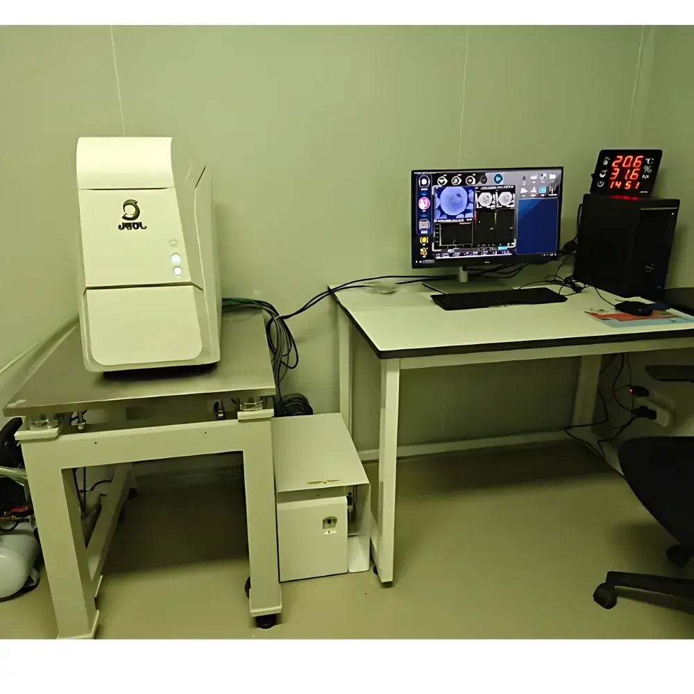
The NIKON JCM-7000 Neoscope Desktop Scanning Electron Microscope (SEM) is an engineered solution for laboratories requiring high-resolution surface imaging without the infrastructure demands of conventional floor-standing systems. Based on thermionic tungsten-filament electron optics and optimized for ease-of-use in non-dedicated microscopy spaces, the JCM-7000 operates under both high-vacuum and low-vacuum conditions—enabling direct observation of non-conductive or hydrated specimens with minimal or no metal coating. Its core imaging principle relies on raster-scanned electron beam interaction with sample surfaces, generating secondary electrons (SE) for topographic contrast and backscattered electrons (BSE) for atomic-number-dependent compositional contrast. Designed for integration into QC labs, forensic units, materials engineering departments, and academic teaching facilities, the system delivers reproducible 20 nm resolution at standard accelerating voltages (5–15 kV), supporting routine morphological analysis, defect identification, particle sizing, and cross-sectional evaluation across diverse industrial sectors—including pharmaceuticals, electronics, polymers, and metallurgy.
Key Features
- Compact desktop architecture with footprint under 0.5 m²—requires no dedicated shielding room, external cooling water, or uninterruptible power supply
- Dual-mode vacuum system featuring a turbomolecular pump (for high-vacuum operation) and rotary vane pump (for low-vacuum mode), enabling rapid mode switching and stable pressure control down to 10⁻³ Pa
- Intuitive 10.4-inch resistive touchscreen interface with real-time dual-channel display—simultaneously rendering SE and BSE images side-by-side or overlaid for correlative interpretation
- Automated acquisition suite including auto-focus, auto-stigmation, auto-contrast/brightness, and automatic electron gun alignment—reducing operator dependency and minimizing training time
- Motorized precision stage with full-centering capability and ±40 mm X/Y travel range, compatible with standard stubs and custom holders up to Ø80 mm × 50 mm height
- Adjustable electron beam current (Power Current) with four discrete settings to optimize signal-to-noise ratio across varying sample conductivity and topography
- Real-time 3D surface reconstruction algorithm integrated into acquisition software—generating depth maps and pseudo-3D visualizations directly from sequential focus-series acquisitions
Sample Compatibility & Compliance
The JCM-7000 accommodates a broad spectrum of specimen types—from conductive metals and ceramics to insulating polymers, biological tissues, and forensic evidence (e.g., fibers, gunshot residue, toolmarks). In low-vacuum mode, samples with moderate outgassing or limited conductivity can be imaged without sputter-coating, preserving native surface chemistry. For elemental analysis readiness, the chamber includes standardized ports for optional EDS detector integration (compatible with major third-party spectrometers). The system complies with IEC 61000-6-3 (EMC emission standards) and IEC 61000-6-2 (immunity requirements), and its operational protocols align with GLP documentation practices—including timestamped image metadata, user-access logs, and audit-trail-capable session records when paired with compliant network storage. While not certified to FDA 21 CFR Part 11 out-of-the-box, the platform supports secure user authentication and configurable electronic signatures via optional enterprise software modules.
Software & Data Management
NIKON’s Neoscope Control Software (v4.x) provides a unified environment for instrument control, image acquisition, measurement annotation, and report generation. All acquired images embed EXIF-style metadata—including kV, working distance, dwell time, magnification, detector type, vacuum mode, and stage coordinates. Export formats include TIFF (16-bit grayscale), JPEG, PNG, and proprietary .NSD files retaining full acquisition parameters. Batch processing tools support automated scale calibration, particle counting (ISO 13322-1 compliant workflows), and line-profile extraction. Data integrity is reinforced through local encrypted database logging, with optional synchronization to network-attached storage or LIMS platforms via standard SFTP or HTTP APIs. Software updates are delivered through NIKON’s secure firmware portal and validated against ISO/IEC 17025 traceability requirements for metrological software.
Applications
- Quality assurance in automotive component manufacturing—detecting microcracks, porosity, and coating delamination on brake pads, piston rings, and battery electrodes
- Pharmaceutical solid dosage form characterization—assessing granule morphology, tablet surface erosion, and API distribution in excipient matrices
- Forensic trace evidence analysis—distinguishing textile fiber subtypes, comparing paint layer sequences, and identifying wear patterns on toolmarks
- Failure analysis in printed circuit boards—locating solder joint voids, intermetallic compound growth, and dendritic silver migration
- Academic instruction in materials science—teaching electron-specimen interactions, diffraction contrast mechanisms, and quantitative stereology principles using live classroom demonstrations
- Biological sample screening—imaging freeze-dried pollen, insect cuticles, and mineralized tissue sections with minimal preparation artifacts
FAQ
Is the JCM-7000 suitable for uncoated biological samples?
Yes—low-vacuum mode permits imaging of non-conductive, hydrated, or outgassing specimens without sputter coating, though dehydration or critical-point drying is recommended for optimal structural fidelity.
Can EDS elemental analysis be performed on this system?
The JCM-7000 features standardized flanges and electrical interfaces for retrofitting with commercially available silicon drift detector (SDD)-based EDS systems; however, EDS hardware is not included as standard equipment.
What is the typical pump-down time from atmospheric pressure to high vacuum?
Approximately 3–5 minutes under standard ambient conditions (23 °C, 45 % RH), depending on chamber loading and initial moisture content of the sample.
Does the system support automated particle analysis?
Yes—integrated measurement tools enable threshold-based particle detection, size distribution histograms (log-normal or Rosin-Rammler fitting), and shape factor quantification (aspect ratio, circularity, convexity) per ISO 9276-2 guidelines.
Is remote operation supported?
Remote desktop access is enabled via standard VNC or RDP protocols when deployed on a secured institutional network; firewall configuration and role-based access controls must be implemented per organizational IT policy.


