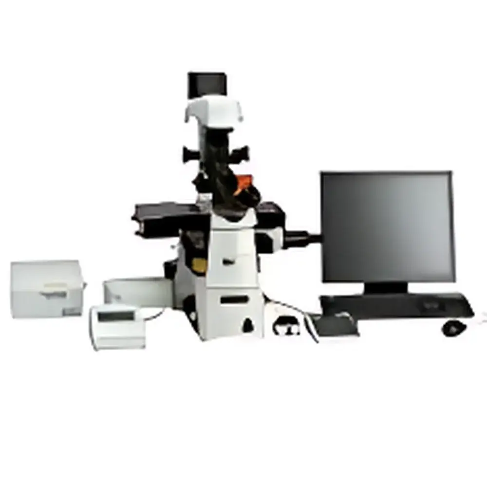
Nikon C1/C1si Laser Confocal Microscope
| Brand | NIKON |
|---|---|
| Origin | Japan |
| Model | C1/C1si |
| Category | Laser Scanning Confocal Microscope |
| Optical Architecture | Upright/Inverted-Compatible Modular Scanning Head |
| Excitation Wavelength Range | 400–650 nm (with multi-laser AOTF control) |
| Pinhole Control | Motorized 4-position aperture wheel |
| Scan Modes | Bidirectional, Rotational (X/Y), Variable-interval Time-Lapse |
| Fluorescence Channels | Simultaneous 3-channel detection (optional DIC integration) |
| TIRF Compatibility | Yes, with Nikon CFI Apo TIRF 60×/100× NA 1.49 objectives |
| FRAP/FLIP/iFRAP Support | Software-enabled via dedicated photobleaching module |
| Laser Capacity | Up to 4 independent lasers (Ar+, HeNe, diode, DPSS) |
| Compliance | Designed for GLP/GMP-aligned workflows |
Overview
The Nikon C1 and C1si Laser Scanning Confocal Microscopes represent a mature, modular implementation of point-scanning confocal fluorescence imaging based on the principle of spatially resolved optical sectioning. By employing a pinhole-apertured detection path aligned conjugately with the focal plane, these systems reject out-of-focus emission light—enabling high-contrast, high-resolution 3D reconstruction of fluorescently labeled biological specimens. Engineered for precision and reproducibility in core facility and academic research environments, the C1 platform integrates optimized scan optics delivering diffraction-limited resolution down to 400 nm excitation, with minimal spherical and chromatic aberration across visible wavelengths. Its compact, pre-aligned scanning head—among the smallest and lightest in its class—is designed for seamless integration with Nikon Eclipse upright and inverted microscope stands, supporting both fixed and live-cell imaging modalities.
Key Features
- Modular, Pre-Calibrated Architecture: All optical modules—including scan head, detector assembly, and laser coupling optics—are factory-aligned and mechanically interlocked, eliminating field recalibration during expansion or maintenance.
- Bidirectional & Rotational Scanning: Bidirectional galvanometer scanning doubles frame rate without compromising linearity; rotational scanning enables angular reorientation of the field of view—critical for longitudinal neural or tissue imaging without mechanical stage rotation.
- Motorized 4-Position Pinhole Wheel: Electronically controlled pinhole selection allows real-time optimization between axial resolution (smaller pinhole) and signal-to-noise ratio (larger pinhole), adapting dynamically to specimen thickness and fluorophore brightness.
- Multi-Laser AOTF Integration: Acousto-optic tunable filter (AOTF) provides millisecond-scale, software-controlled intensity modulation for up to four independent lasers (e.g., 488 nm Ar+, 543 nm HeNe, 638 nm diode, 561 nm DPSS), ensuring precise photostimulation and multicolor excitation balance.
- TIRF-Ready Platform: Native compatibility with Nikon’s CFI Apo TIRF 60× and 100× oil immersion objectives (NA 1.49)—the highest numerical aperture available for total internal reflection fluorescence—enables evanescent wave excitation within ~100 nm of the coverslip surface.
Sample Compatibility & Compliance
The C1/C1si accommodates standard 24 × 60 mm and 35 mm glass-bottom dishes, multi-well plates (6–96-well), and custom chambered slides. It supports live-cell imaging under environmental control (temperature, CO₂, humidity) when paired with compatible incubation chambers. From a regulatory standpoint, the system is engineered to support workflows compliant with ISO/IEC 17025 (for calibration traceability), ASTM E2877-13 (microscopy performance verification), and USP (fluorescence microscopy validation). When operated with Nikon NIS-Elements AR software configured for audit trail logging, data acquisition meets FDA 21 CFR Part 11 requirements for electronic records and signatures in regulated environments.
Software & Data Management
NIS-Elements AR (Advanced Research) serves as the unified acquisition and analysis environment. It provides full hardware control—including AOTF, pinhole, PMT gain/voltage, Z-stack parameters, and time-lapse intervals—with scriptable automation via Python and MATLAB APIs. The software natively supports FRAP, FLIP, and iFRAP protocols through an intuitive region-of-interest (ROI) editor that accepts arbitrary polygonal, annular, or hollow geometries. All acquisition metadata—including laser power history, pinhole position, objective ID, and environmental sensor logs—are embedded in TIFF and ND2 file headers. Raw data export adheres to open formats (OME-TIFF) for interoperability with ImageJ/Fiji, Imaris, and Huygens deconvolution platforms.
Applications
The C1/C1si is routinely deployed in quantitative cell biology for dynamic studies including intracellular protein trafficking (e.g., GFP-tagged kinesin motility), membrane receptor diffusion (via FRAP), organelle morphology dynamics (mitochondrial fission/fusion), and sub-diffraction spatial mapping of synaptic proteins using structured illumination-compatible acquisition modes. In developmental biology, it supports long-term time-lapse imaging of zebrafish or Drosophila embryos with minimal phototoxicity due to efficient photon collection and low-dose excitation scheduling. Its TIRF integration further enables single-molecule localization and adhesion plaque turnover analysis in adherent mammalian cells.
FAQ
Can the C1/C1si be upgraded from widefield to confocal mode?
Yes—the scanning head mounts directly onto existing Nikon Eclipse stands equipped with appropriate laser ports and detector interfaces; no optical realignment is required.
Is NIS-Elements AR validated for GxP compliance?
When configured with electronic signature, audit trail, and user access controls enabled, NIS-Elements AR supports 21 CFR Part 11 compliance in pharmaceutical and clinical research settings.
What is the maximum achievable Z-resolution in thick tissue sections?
Axial resolution depends on objective NA, pinhole size, and refractive index matching; with the CFI Plan Apo λ 60×/1.40 oil objective and 1 Airy unit pinhole, typical Z-step precision is ≤0.5 µm in fixed, cleared samples.
Does the system support spectral unmixing?
No—the C1/C1si uses conventional bandpass-filtered PMT detection; spectral unmixing requires the Nikon A1R+ or Ni-E platforms with spectral detectors.
Are service contracts and extended calibration options available internationally?
Yes—NIKON Metrology and authorized service partners provide annual preventive maintenance, wavelength calibration verification, and ISO 17025-compliant intensity calibration reports upon request.


