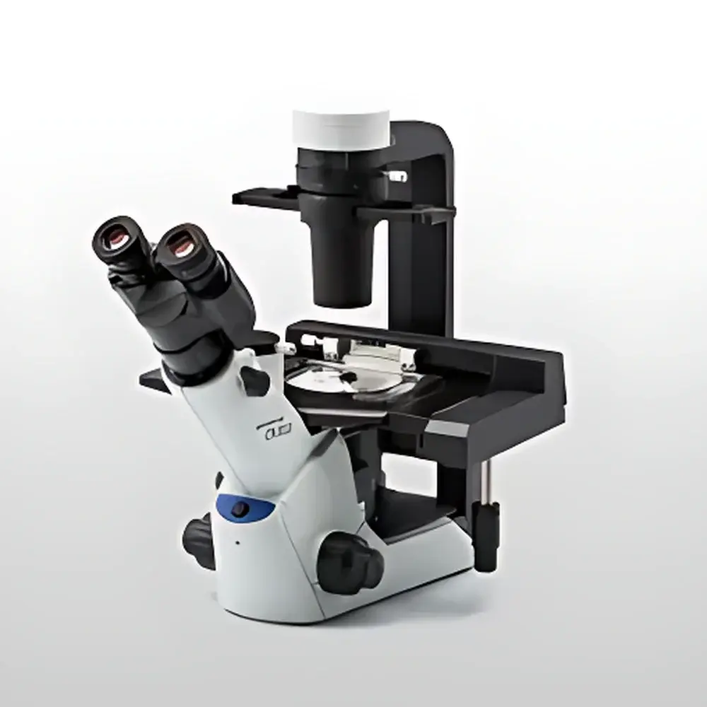
Olympus CKX53 Inverted Microscope
| Brand | Olympus |
|---|---|
| Origin | Japan |
| Manufacturer | Olympus Corporation |
| Product Type | Imported Instrument |
| Model | CKX53 |
| Microscope Configuration | Trinocular |
| Category | Inverted Biological Microscope |
| Primary Application | Live-Cell Imaging & Cell Culture Monitoring |
| Ergonomic Design | 45° Tilting Eyepiece Tube, Compact Footprint (≈270 × 380 mm), Weight: ~7 kg |
| Optical System | Integrated Phase Contrast (iPC) for 4×, 10×, 20×, 40× Objectives |
| Objective Specifications | PLN2× (22 mm Field of View, 11 mm Annular Aperture, High Contrast), PLCN4× (Long Working Distance, ≥19 mm, Compatible with Multilayer Culture Vessels up to 190 mm Height) |
| Sterilization Compatibility | UV-resistant Coating Enables Placement Inside Laminar Flow Hoods |
| Stage Flexibility | ±70 mm Lateral Extension, Removable Condenser, Universal Adapter Support for Petri Dishes (up to three 35 mm), Microplates (no adapter required), and T-flasks |
Overview
The Olympus CKX53 is a purpose-engineered inverted microscope designed specifically for routine cell culture monitoring, live-cell observation, and aseptic handling in biosafety cabinets and laminar flow hoods. Its optical architecture leverages Olympus’ proprietary Integrated Phase Contrast (iPC) system—eliminating the need for manual alignment or replacement of annular apertures across 4×, 10×, 20×, and 40× magnifications. This ensures rapid, reproducible contrast enhancement for unstained, transparent specimens such as adherent mammalian cells, organoids, and primary cultures—without introducing artifacts associated with staining or fixation. The CKX53 employs a finite-conjugate optical path optimized for stability under variable environmental conditions typical of tissue culture facilities, including ambient temperature fluctuations and vibration-prone benchtops. Its compact mechanical design (270 × 380 mm footprint, ~7 kg mass) enables deployment directly inside Class II biosafety cabinets, while its UV-resistant surface coating permits routine decontamination via 254 nm ultraviolet irradiation without optical degradation.
Key Features
- Trinocular optical head with 45° inclined eyepieces and ergonomic butterfly-shaped tube—supports both seated and standing postures while minimizing cervical strain during extended observation sessions.
- Integrated Phase Contrast (iPC) system enabling immediate high-contrast imaging at 4×, 10×, 20×, and 40× magnifications—no centering tools or aperture swaps required.
- PLN2× objective delivering a 22 mm field of view and optimized contrast for rapid confluence assessment and colony selection; ideal for low-magnification screening of heterogeneous cultures.
- PLCN4× objective featuring ≥19 mm working distance—designed to accommodate multilayer cell culture vessels (e.g., triple-layer flasks) up to 190 mm in height without stage interference.
- Modular stage system with ±70 mm lateral extension, removable Abbe condenser, and universal adapter compatibility—including direct placement of 6-/12-/24-/96-well microplates, 35 mm Petri dishes (up to three simultaneously), and T-25/T-75 flasks.
- Human-centered control layout: coaxial coarse/fine focus knobs, light intensity dial, and beam-splitter selector positioned within natural hand reach—reducing repetitive motion stress during daily QC checks or subculturing workflows.
Sample Compatibility & Compliance
The CKX53 accommodates standard cell culture formats without modification: sterile plasticware (e.g., Corning, Falcon), glass-bottom dishes, and silicone-based microfluidic chambers. Its open-stage architecture supports real-time manipulation using micropipettes or patch-clamp rigs adjacent to the objective. The instrument complies with ISO 13485–aligned manufacturing standards and meets IEC 61000-6-3 (EMC emission) and IEC 61000-6-2 (immunity) requirements. While not FDA-cleared as a diagnostic device, its optical repeatability and mechanical stability support GLP-compliant documentation of cell morphology changes in preclinical assay development per OECD TG 442D and ASTM E2929-13 guidelines.
Software & Data Management
The CKX53 is compatible with Olympus cellSens Entry and cellSens Standard software suites (sold separately), enabling time-lapse acquisition, multi-channel fluorescence overlay (when equipped with optional filter cubes), region-of-interest (ROI) intensity profiling, and automated confluence quantification. All captured images embed EXIF metadata—including objective ID, magnification, exposure time, and illumination intensity—for audit-trail integrity. When integrated with networked storage systems, image datasets adhere to DICOM Supplement 145 (Microscopy) conventions, facilitating interoperability with LIMS platforms compliant with 21 CFR Part 11 electronic record requirements.
Applications
- Routine passage validation and morphology assessment of adherent cell lines (e.g., HeLa, HEK293, CHO-K1).
- Monitoring differentiation dynamics in stem cell cultures and 3D spheroid models.
- Pre-screening transfection efficiency prior to downstream assays (e.g., qPCR, Western blot).
- Supporting aseptic procedures—cell harvesting, medium exchange, and cryovial preparation—within laminar flow hoods.
- Time-lapse documentation of wound-healing assays, migration kinetics, and apoptosis progression under physiological conditions.
FAQ
Can the CKX53 be used inside a CO2 incubator?
No—the CKX53 is not rated for humidified, 5% CO2, 37°C environments. It is intended for use in ambient laboratory or biosafety cabinet settings only.
Is fluorescence capability built-in?
Fluorescence observation requires optional filter cubes, LED illumination modules (e.g., U-HGLGPS), and appropriate objectives (e.g., UPLSAPO series); the base CKX53 configuration supports brightfield and phase contrast only.
What is the maximum vessel height supported without stage modification?
With the condenser fully retracted and the universal adapter removed, the CKX53 accommodates vessels up to 190 mm tall—sufficient for commercially available multilayer culture flasks.
Does the iPC system require calibration between objectives?
No—iPC alignment is factory-optimized and mechanically locked per objective turret position; no user recalibration is necessary across the 4×–40× range.
Is the microscope compatible with third-party digital cameras?
Yes—via C-mount (0.5× reduction lens included) or trinocular port with standard 23.2 mm thread; supported by common machine vision SDKs (e.g., Thorlabs DCx, FLIR Spinnaker).


