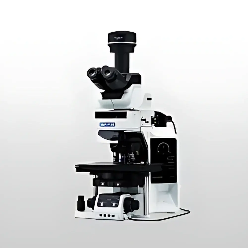
Olympus BX63 Fully Motorized Upright Fluorescence Microscope
| Brand | Olympus |
|---|---|
| Origin | Japan |
| Manufacturer Type | Original Equipment Manufacturer (OEM) |
| Origin Category | Imported Instrument |
| Model | BX63 |
| Instrument Type | Upright Fluorescence Microscope |
| Instrument Class | Research-Grade Fluorescence Microscope |
| Objective Turret | Motorized |
| Focus Mechanism | Manual and Motorized |
| Condenser Control | Manual and Motorized |
| Observation Head | Binocular and Trinocular |
| DIC Compatibility | Integrated Differential Interference Contrast (DIC) Module Available |
| Stage | Manual, Mechanical, and Ultrasonic Piezo Motorized XY Stage |
| Fluorescence Filter Sets | Multiple Standard and Customizable Excitation/Emission Filter Combinations |
| Control Interface | Touchscreen Controller with Preset Recall and Workflow Manager |
Overview
The Olympus BX63 is a research-grade, fully motorized upright fluorescence microscope engineered for high-precision live-cell imaging, multi-channel fluorescence quantification, and reproducible structural analysis in life science laboratories. Built upon Olympus’ proven UIS2 optical platform, the BX63 employs Köhler illumination geometry and high-transmission, apochromatic fluorite objectives to ensure exceptional signal-to-noise ratio, minimal photobleaching, and chromatic fidelity across UV–visible spectral bands (340–780 nm). Its core architecture integrates synchronized motorization of the objective turret, Z-axis focus drive, XY piezoelectric stage, condenser height and aperture control, and fluorescence filter cube changer—enabling full hardware-level coordination under software command. This level of integration supports rigorous experimental protocols requiring temporal stability, positional repeatability, and compliance with GLP/GMP documentation workflows.
Key Features
- Fully synchronized motorization: Independent yet coordinated control of objective turret (6–7 position), fine/coarse Z-focus (10 nm step resolution), ultrasonic piezo-driven XY stage (±5 mm travel, 50 nm repeatability), and motorized condenser (height and iris).
- Dual-mode operation: Seamless switching between manual ergonomics and automated acquisition via detachable touchscreen controller—designed for benchtop flexibility without sacrificing precision.
- Integrated DIC capability: Optional Nomarski-style differential interference contrast module compatible with all UIS2 objectives, enabling label-free phase contrast imaging alongside fluorescence channels.
- Modular fluorescence optics: Standard support for up to 6 filter cubes (e.g., DAPI/FITC/TRITC/Cy5), with optional motorized cube changer and LED or metal-halide light source integration (e.g., X-Cite 120LED or MT20G).
- Workflow Manager in cellSens software: Enables scripted multi-position, multi-channel, Z-stack, and time-lapse acquisitions—including conditional triggering, autofocus correction per field, and hardware-synchronized exposure timing.
- Regulatory-ready architecture: Supports audit trail logging, user access levels, electronic signatures, and metadata embedding compliant with FDA 21 CFR Part 11 requirements when deployed with validated cellSens Digital Imaging Software v2.4 or later.
Sample Compatibility & Compliance
The BX63 accommodates standard glass slides (1–3 mm thickness), petri dishes (35–100 mm), multi-well plates (6–96-well), and custom chambered coverslips for live-cell perfusion studies. Its rigid cast-aluminum frame and fixed-stage design minimize mechanical drift during long-term acquisitions (>12 h), while the motorized stage maintains sub-micron positional fidelity across thermal gradients (20–25 °C ambient). The system complies with IEC 61000-6-3 (EMC emission standards), IEC 61010-1 (safety for laboratory equipment), and ISO 13485–aligned manufacturing quality systems. All optical components meet JIS B 7151 specifications for transmission uniformity and wavefront error.
Software & Data Management
Olympus cellSens Digital Imaging Software serves as the unified interface for instrument control, image acquisition, quantitative analysis, and metadata management. It provides real-time hardware feedback, ROI-based intensity profiling, spectral unmixing (with linear unmixing algorithm), and batch processing pipelines exportable to TIFF, OME-TIFF, or HDF5 formats. Audit trail functionality logs operator ID, timestamp, parameter changes, and image modifications—fully traceable for regulatory submissions. Integration with third-party platforms (e.g., ImageJ/Fiji via Bio-Formats, MATLAB via COM/ActiveX, or Python via PyMicroscope SDK) enables extensible workflow development.
Applications
- High-content screening (HCS) of fixed and live mammalian cells using multiplexed immunofluorescence labeling.
- Subcellular organelle dynamics tracking (e.g., mitochondria fission/fusion, lysosomal pH mapping) with ratiometric dyes and time-lapse Z-stacks.
- Neuronal morphology reconstruction in brain slice cultures using DiI/DiO staining and confocal-compatible widefield deconvolution.
- Quantitative colocalization analysis (Pearson’s coefficient, Mander’s overlap) in developmental biology specimens.
- Standardized histopathology review with DIC–fluorescence correlation on FFPE tissue sections.
- GMP-compliant QC of bioprocess-derived cell therapies via morphometric and fluorescence intensity validation protocols.
FAQ
Does the BX63 support automated Z-stack acquisition with hardware autofocus?
Yes—the BX63 integrates a motorized Z-drive with optional hardware autofocus sensor (ZDC2), enabling closed-loop focus maintenance during multi-hour time-lapse or large-area tiled acquisitions.
Can the touchscreen controller be used remotely or mounted off the microscope frame?
Yes—the detachable controller features magnetic mounting brackets and supports USB-C tethering up to 3 m; it also operates wirelessly via optional Wi-Fi dongle for remote benchtop positioning.
Is DIC functionality available natively or as an add-on module?
DIC is offered as a factory-installed or field-upgradeable option, including Wollaston prisms, strain-free condenser, and matching Nomarski-compatible objectives (e.g., UPLSAPO 40x/0.95).
What fluorescence light sources are compatible with the BX63?
The system accepts mercury arc (U-LH100HGAPO), metal halide (MT20G), and solid-state LED sources (X-Cite 120LED, SOLA SE), all controllable via TTL or analog voltage signals from cellSens.
How does the BX63 ensure data integrity for regulated environments?
Through cellSens’ Part 11-compliant mode: electronic signatures, role-based permissions, immutable audit trails, and encrypted raw data containers (OME-TIFF with embedded EXIF and OMEXML metadata).


