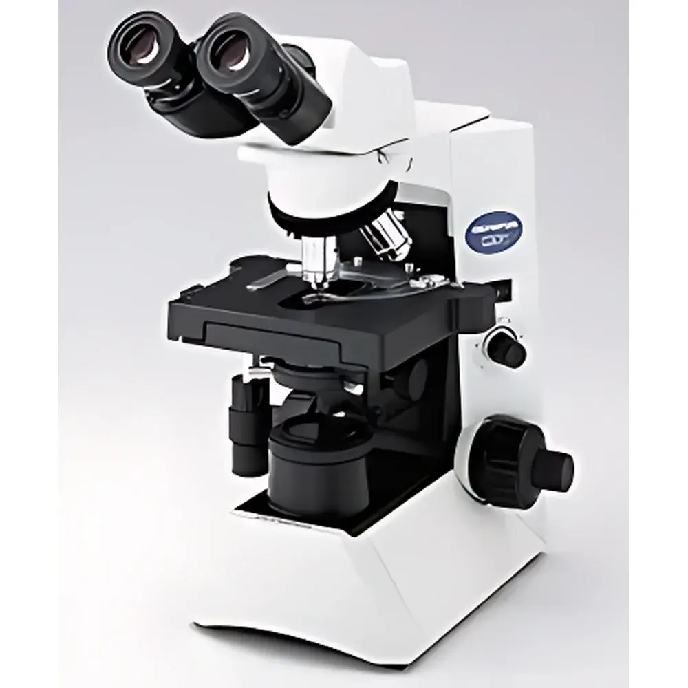
Olympus CX31 Biological Microscope
| Brand | Olympus |
|---|---|
| Origin | Japan |
| Model | CX31 |
| Optical System | UIS (Infinity-Corrected) |
| Illumination | Built-in Köhler-transmitted illumination with 6V30W halogen lamp (100–120 V / 220–240 V, 0.85/0.45 A, 50/60 Hz) |
| Focusing Mechanism | Coaxial coarse/fine focus |
| Nosepiece | Fixed inward-sloping 4-position objective turret |
| Eyepiece Tube | Binocular or trinocular |
| Stage | Mechanical stage, 188 × 134 mm |
| Condenser | Abbe condenser with built-in daylight filter and iris diaphragm |
| Dimensions & Weight | 233 × 411 × 367.5 mm (W × H × D) |
Overview
The Olympus CX31 Biological Microscope is a robust, entry-to-mid-tier upright microscope engineered for routine brightfield observation in academic, clinical, and industrial life science laboratories. Designed around Olympus’ UIS (Universal Infinity System) optical architecture, the CX31 delivers high-contrast, aberration-corrected imaging across the visible spectrum—enabling consistent resolution and color fidelity essential for morphological assessment of unstained and stained biological specimens. Its fixed 4-objective turret, precision mechanical stage, and integrated Köhler illumination system provide stable, reproducible imaging conditions ideal for prolonged daily use in teaching labs, quality control workflows, and basic research applications involving live or fixed cells, microorganisms, tissue sections, and suspension cultures.
Key Features
- UIS infinity-corrected optical system ensures optimal image flatness and chromatic correction, compatible with standard 4×, 10×, 40×, and 100× oil-immersion objectives (all sold separately or as optional kits)
- Integrated Köhler illumination with 6V30W halogen lamp supports uniform, glare-free illumination—adjustable via centerable field and aperture diaphragms for precise contrast control
- Coaxial coarse/fine focusing mechanism offers ergonomic operation: 36.8 mm coarse travel per revolution, 25 mm total vertical range, and 0.2 mm fine focus increment with adjustable tension and mechanical coarse-stop limiters
- Sturdy cast-metal frame and vibration-damped base ensure mechanical stability during extended observation or manual documentation
- Binocular or trinocular viewing head options support both direct visual examination and simultaneous imaging via C-mount or third-party camera adapters (e.g., USB3.0 or HDMI-capable digital eyepieces)
- Abbe condenser (NA 1.25) with built-in daylight filter and iris diaphragm enables optimal numerical aperture matching for all dry and oil-immersion objectives
Sample Compatibility & Compliance
The CX31 accommodates standard 1″ × 3″ (25 × 75 mm) glass slides and coverslips (0.13–0.17 mm thickness), supporting routine histology, hematology, microbiology, and cytology preparations. It is routinely deployed in GLP-aligned educational labs and QC environments where traceability and procedural consistency are required—notably in undergraduate biology instruction, pharmaceutical raw material screening, and environmental microbial analysis. While the CX31 itself does not carry FDA 510(k) clearance or CE-IVD designation, its optical performance complies with ISO 8578:2017 (microscopes — requirements and tests) and supports adherence to ASTM E2919-21 (standard guide for selection and use of optical microscopes in materials evaluation). Its mechanical design meets IEC 61010-1 safety standards for laboratory electrical equipment.
Software & Data Management
As a hardware-only platform, the CX31 does not include proprietary acquisition software. However, its trinocular port is mechanically and optically calibrated for third-party digital imaging solutions—including Olympus cellSens Entry (discontinued but still supported), Thorlabs DCx series drivers, or open-source platforms such as Micro-Manager v2.x. When paired with compliant USB3.0 cameras, the system supports time-lapse capture of dynamic processes (e.g., mitotic progression in adherent cell lines or motility assays), with metadata logging (timestamp, magnification, exposure) enabled through host software. Audit trails, user access logs, and electronic signatures require integration with external LIMS or ELN systems—no native 21 CFR Part 11 functionality is embedded.
Applications
- Cell morphology and viability assessment in mammalian, insect, and plant cell cultures
- Microbial identification and quantification in clinical microbiology and food safety testing
- Routine hematology (e.g., peripheral blood smear evaluation) and parasitology (e.g., malaria parasite detection)
- Basic immunohistochemistry and H&E-stained tissue section screening
- Industrial QC of fermentation broths, yeast suspensions, and spore preparations
- Undergraduate laboratory instruction in genetics, developmental biology, and microbiology curricula
FAQ
Is the CX31 suitable for fluorescence microscopy?
No—the CX31 lacks epi-illumination capability, filter cubes, and dichroic mirrors required for fluorescence. For fluorescence applications, consider the Olympus BX43 or IX53 series.
Can I upgrade the CX31 to accommodate phase contrast?
Yes—phase contrast is supported via optional phase contrast condenser (U-PCF), phase objectives (e.g., UPLFLN-PH series), and centerable alignment telescopes. Installation requires mechanical alignment but no optical modification to the base unit.
What is the maximum usable magnification with the standard CX31 configuration?
With a 10× eyepiece and 100× oil-immersion objective (NA 1.25), theoretical resolution is ~0.22 µm (using λ = 550 nm), yielding effective usable magnification up to 1000×. Higher nominal magnifications introduce empty magnification without added detail.
Does Olympus provide service documentation or calibration certificates for the CX31?
Olympus provides optical alignment procedures and maintenance guides under NDA for authorized service partners. Factory calibration certificates are not issued for standard CX31 units; users requiring metrological traceability must engage third-party accredited labs for verification per ISO/IEC 17025.
Is the CX31 compatible with digital pathology scanning workflows?
Not natively—the CX31 lacks motorized stage, Z-stack automation, or whole-slide scanning optics. It may serve as a validation reference instrument alongside dedicated slide scanners (e.g., Hamamatsu NanoZoomer or Leica GT 450), but cannot replace them in diagnostic archival pipelines.


