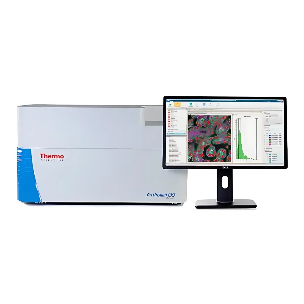
Thermo Scientific CellInsight CX7 High-Content Screening and Analysis Platform
| Origin | USA |
|---|---|
| Manufacturer Type | Authorized Distributor |
| Origin Category | Imported |
| Model | CellInsight CX7 |
| Price Range | USD 420,000 – 490,000 (FOB) |
| Temperature Control | Not Integrated |
| Humidity Control | Not Integrated |
| Throughput Capacity | Up to 1,000 wells/hour (typical, dependent on assay configuration and imaging mode) |
Overview
The Thermo Scientific™ CellInsight™ CX7 High-Content Screening (HCS) and Analysis Platform is a fully integrated, benchtop-grade automated imaging system engineered for quantitative, multi-parametric cellular phenotyping in life science research and early-stage drug discovery. Leveraging advanced widefield and spinning-disk confocal microscopy architectures, the platform delivers high-fidelity subcellular resolution across diverse sample formats—including adherent and suspension cells, 3D spheroids, tissue sections, and organoids—without requiring user intervention in focus calibration or illumination optimization. Its core measurement principle combines multi-channel fluorescence and color brightfield imaging with real-time laser-based autofocus, enabling consistent z-stack acquisition even across non-uniform well distributions (e.g., edge effects in 384- or 1536-well plates). The system’s optical architecture supports both kinetic and endpoint assays, with precise temporal control over exposure, illumination intensity, and channel sequencing—critical for minimizing phototoxicity and ensuring reproducibility in longitudinal studies.
Key Features
- Seven-channel solid-state LED light engine providing stable, tunable excitation across 365–770 nm—enabling robust multiplexing with standard fluorophores (e.g., DAPI, FITC, TRITC, Cy5) and H&E-compatible chromogenic detection
- Spinning-disk confocal module with dual-disk configuration for enhanced optical sectioning of thick specimens (up to 100 µm), delivering improved signal-to-noise ratio and reduced out-of-focus blur in 3D matrices
- Cooling-controlled scientific-grade CCD camera with 16-bit dynamic range and <0.5 e⁻ read noise—optimized for low-light fluorescence quantification and high-contrast brightfield morphology analysis
- Patented laser autofocus system operating in real time per field-of-view, eliminating reliance on reference wells or pre-scan Z-maps and reducing total acquisition time by up to 40% versus conventional motorized focus methods
- Modular hardware design supporting integration with third-party liquid handlers and robotic arms (via standard SLAS/ANSI-compliant plate interfaces), compliant with HTS workflow standards
- Pre-aligned optical path with factory-calibrated chromatic correction—ensuring pixel-level registration across all fluorescence and brightfield channels without post-acquisition alignment
Sample Compatibility & Compliance
The CellInsight CX7 accommodates standard microplate formats (6–1536-well), glass-bottom dishes, chamber slides, and histological tissue sections mounted on standard microscope slides. It supports fixed and live-cell assays, including pH- and Ca²⁺-sensitive probes, mitochondrial membrane potential dyes, and viability markers compatible with FDA-recommended labeling protocols (e.g., USP , ISO 13485 Annex A for IVD-related validation). All imaging parameters—including exposure time, gain, LED intensity, and z-step interval—are logged with timestamped metadata in accordance with 21 CFR Part 11 requirements when used with validated HCS Studio software configurations. System qualification documentation (IQ/OQ/PQ templates) and audit trail functionality support GLP and GMP-aligned workflows in regulated environments.
Software & Data Management
HCS Studio™ Cell Analysis Software serves as the unified analytical environment, offering two operational tiers: guided workflow mode for novice users and scriptable analysis builder for advanced developers. Over 30 pre-validated application modules—including apoptosis (Annexin V/PI), autophagy (LC3 puncta), cell cycle (EdU/DNA), DNA damage (γH2AX foci), neurite outgrowth, and synapse density quantification—are provided with parameter presets optimized for common cell lines (e.g., HeLa, U2OS, iPSC-derived neurons). Each module includes built-in background subtraction, adaptive thresholding, subcellular object segmentation (nucleus/cytoplasm/membrane), and statistical reporting (mean intensity, count, texture, shape descriptors). For custom assay development, users can chain image processing steps via drag-and-drop nodes, adjust sensitivity thresholds in real time, and export analysis pipelines as reusable .xml protocols. All raw images, processed data, and metadata are stored in an ACID-compliant SQLite database with optional network drive mirroring; results export directly to CSV, Excel, or Cytobank-compatible FCS format—no offline post-processing required.
Applications
The platform is routinely deployed in target identification and validation, mechanism-of-action studies, toxicology profiling (e.g., mitochondrial dysfunction, lysosomal accumulation), and phenotypic screening of CRISPR/Cas9 knockout libraries. Its ability to resolve spatial heterogeneity within single cells makes it particularly valuable for immuno-oncology applications—such as quantifying PD-L1 membrane localization alongside T-cell infiltration markers in co-culture models—or for neurodegenerative disease modeling where axonal transport kinetics and synaptic vesicle distribution require multi-z, multi-channel correlation. In translational pathology, the simultaneous acquisition of H&E-like brightfield morphology and immunofluorescence enables direct cross-modality registration—facilitating digital biomarker discovery in FFPE tissue microarrays under CLIA-certified laboratory conditions.
FAQ
Does the CellInsight CX7 support live-cell imaging over extended durations?
Yes—the system includes environmental enclosure options (sold separately) that integrate CO₂ regulation, temperature stabilization (37 °C ±0.5 °C), and humidity control for kinetic assays up to 72 hours.
Can HCS Studio software be validated for use in FDA-submitted studies?
Yes—when deployed in locked-down, audit-trail-enabled configurations with electronic signatures and change control logs, HCS Studio meets ALCOA+ principles and supports submission-ready documentation packages.
Is the spinning-disk confocal module interchangeable with widefield optics?
No—the optical train is permanently aligned at the factory; however, users select imaging mode (widefield vs. confocal) dynamically per acquisition protocol without mechanical reconfiguration.
What file formats does the system natively export for downstream analysis?
Raw data is saved in OME-TIFF format with embedded metadata; processed results export to CSV, XLSX, and FCS 3.1 for flow cytometry-style gating. Custom Python/R plugins can be loaded via the HCS Studio SDK.
How is instrument performance monitored over time?
The platform includes daily QC routines—automated LED intensity calibration, autofocus precision verification, and camera dark-current measurement—with trend reports accessible via the web-based system dashboard.


