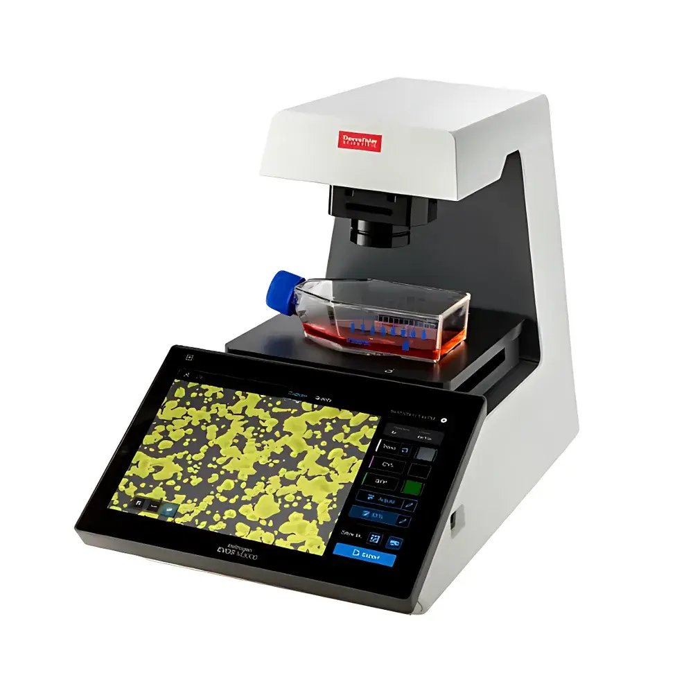
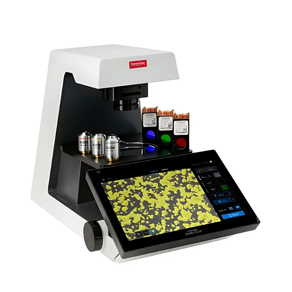
Invitrogen EVOS M3000 Digital Smart Cell Imaging System
| Brand | Invitrogen |
|---|---|
| Origin | USA |
| Manufacturer Type | Original Equipment Manufacturer (OEM) |
| Product Category | Imported Instrument |
| Model | EVOS M3000 |
| Instrument Type | Inverted Microscope |
| Imaging Modes | Brightfield, Phase Contrast, Color, Fluorescence |
| Camera | High-Stability CMOS Sensor |
| Software | Embedded AI-Powered Cell Confluence Analysis Engine |
| Connectivity | Ethernet/Wi-Fi Enabled for Cloud Integration |
| Dimensions | Compact Footprint Optimized for Biosafety Cabinets and CO₂ Incubators |
| Objective Compatibility | EVOS™ Standard Objectives (1.25×–60×) and Filter Cubes |
Overview
The Invitrogen EVOS M3000 Digital Smart Cell Imaging System is a fully integrated, inverted digital microscope engineered for routine, high-reproducibility cell culture monitoring in academic, pharmaceutical, and biomanufacturing laboratories. It operates on a dual-principle architecture: optical imaging via precision-corrected optics combined with embedded computational image analysis grounded in validated machine learning models. Unlike conventional microscopes requiring post-acquisition software or manual thresholding, the EVOS M3000 performs real-time confluence quantification directly from live optical feed—without saving intermediate images—thereby eliminating observer bias, reducing inter-operator variability, and aligning with Good Laboratory Practice (GLP) documentation requirements. Its optical path supports four native modalities—brightfield, phase contrast, color (RGB), and fluorescence—enabling longitudinal assessment of morphology, viability, transfection efficiency, and reporter expression without hardware reconfiguration.
Key Features
- Sub-second confluence analytics: Proprietary on-device AI model delivers quantitative confluence percentage per field-of-view in <1 second, validated against standard hemocytometer and automated image segmentation benchmarks (e.g., NIH ImageJ with Trainable Weka Segmentation).
- No-training workflow: 10.1-inch capacitive touchscreen interface with gesture-driven navigation; all functions—including focus, exposure, mode switching, and analysis initiation—are accessible within two taps.
- Incubator- and biosafety cabinet-compatible form factor: 24.5 × 29.5 × 34.0 cm (W × D × H); weight: 7.8 kg; fanless thermal design ensures stable operation inside 37°C/5% CO₂ environments without condensation risk.
- Modular optical architecture: Interchangeable EVOS light cubes (including DAPI, GFP, RFP, Cy5, and transmitted-light optimized filters) and standardized objective mount accept 1.25× to 60× magnifications with parfocal alignment across modalities.
- Embedded data integrity controls: Automatic timestamping, user ID logging, and audit-trail-enabled analysis history—fully compliant with FDA 21 CFR Part 11 requirements when deployed with optional secure cloud gateway.
Sample Compatibility & Compliance
The EVOS M3000 supports standard tissue culture formats including 6–96-well plates, T-flasks (T25–T225), Petri dishes (35–150 mm), and chamber slides. Its long-working-distance objectives accommodate vessels with bottom thicknesses from 0.15–2.0 mm (including glass-bottom and polymer substrates). All optical components meet ISO 10934-1 (microscope performance standards) and IEC 61000-6-3 (EMC emissions). The system’s firmware and analysis engine have undergone internal verification per ASTM E2577 (Standard Practice for Digital Imaging in Microscopy) and are routinely deployed in GLP-compliant cell banking and QC release testing workflows.
Software & Data Management
The onboard operating system runs a deterministic real-time kernel, ensuring consistent frame capture timing and analysis latency. Raw sensor data (16-bit TIFF stream) and processed confluence metadata (CSV + JSON) are exportable via USB 3.0 or network share. Optional cloud integration enables SFTP-based transfer to LIMS or ELN platforms (e.g., LabArchives, Benchling), with role-based access control and versioned dataset archiving. Audit trails record operator actions, parameter changes, and analysis events—including timestamps, IP addresses (for network sessions), and cryptographic hash values for output files—supporting traceability under ISO/IEC 17025 and USP analytical instrument qualification guidelines.
Applications
- Passage timing optimization in primary and stem cell cultures (e.g., iPSCs, MSCs, hESCs)
- Real-time monitoring of CRISPR-edited clone expansion and phenotypic drift
- Transfection efficiency scoring across plasmid, siRNA, and lentiviral delivery methods
- Pre- and post-thaw viability assessment in cryopreserved biobank inventories
- QC release testing of therapeutic cell products per ICH Q5D and USP
FAQ
Does the EVOS M3000 require external computers or software licenses to perform confluence analysis?
No. All image acquisition, preprocessing, and AI-based confluence computation occur locally on the device’s dedicated processing unit. No subscription, dongle, or third-party license is required.
Can the system be validated for use in GMP-regulated environments?
Yes. The EVOS M3000 supports IQ/OQ/PQ documentation packages, including calibration certificates for focus repeatability (±0.5 µm), exposure linearity (R² > 0.999), and confluence accuracy (±3% absolute deviation vs. reference histology-based ground truth).
Is fluorescence imaging quantitative across multiple channels?
Fluorescence intensity is captured in linear 16-bit dynamic range per channel; however, absolute quantification requires separate photometric calibration using NIST-traceable fluorescent standards—supported via optional calibration module.
How is data security handled during cloud transmission?
All network transfers use TLS 1.2+ encryption; credentials are stored in hardware-backed secure enclaves; no raw image data is retained on the device after successful upload unless explicitly configured for local backup.


