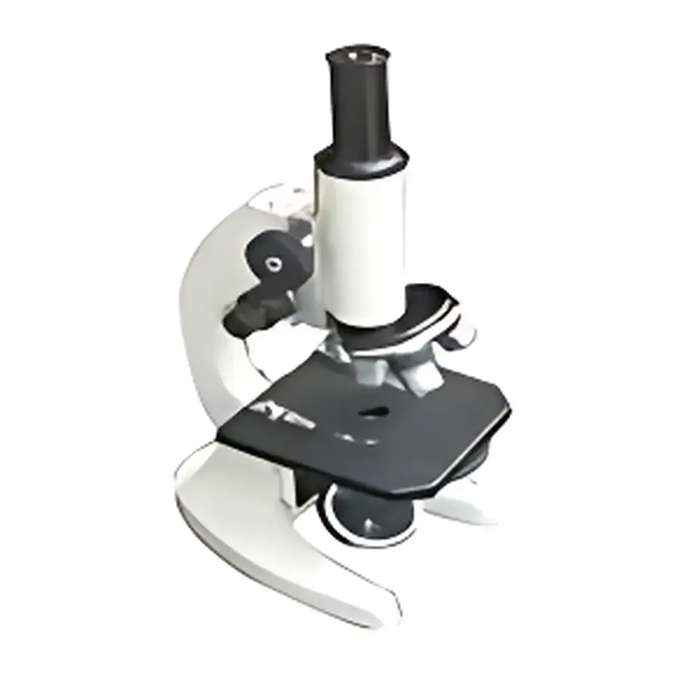
Yoke XSP-1CA Monocular Biological Microscope
| Brand | Yoke |
|---|---|
| Origin | Shanghai, China |
| Manufacturer Type | Manufacturer |
| Country of Origin | China |
| Model | XSP-1CA |
| Price | Upon Request |
| Total Magnification Range | 40×–1600× |
| Eyepiece | WF10×/Φ18 mm, WF16×/Φ11 mm |
| Objective Lenses | 4×/0.10, 10×/0.25, 40×(S)/0.65, 100×(S) oil/1.25 |
| Nosepiece | Quadruple revolving nosepiece |
| Mechanical Tube Length | 160 mm |
| Stage | Dual-layer mechanical stage (125 × 120 mm), travel range 70 × 30 mm |
| Condenser | N.A. 1.25 Abbe condenser with centering adjustment, iris diaphragm and filter holder |
| Focusing Mechanism | Coaxial coarse/fine focus (15 mm travel), fine focus graduation: 0.002 mm |
| Eyepiece Tube | Monocular, inclined at 45°, rotatable 360° |
| Illumination | Reflected natural light (no built-in lamp) |
Overview
The Yoke XSP-1CA is a precision-engineered monocular biological microscope designed for routine clinical and applied life science applications in resource-constrained or entry-level laboratory environments. Built to the classical 160 mm mechanical tube length standard, it operates on conventional brightfield optical principles—relying on transmitted illumination through stained or naturally contrasted biological specimens. Its optical architecture adheres to long-standing international conventions for parfocal, parcentric objective systems, ensuring consistent focus and field centering across magnifications. The instrument is optimized for stability, ergonomic usability, and long-term mechanical repeatability—making it suitable for high-volume manual microscopy tasks such as blood smear analysis, urine sediment examination, parasitology screening, and basic histological evaluation in small clinical labs, veterinary clinics, agricultural extension centers, and aquaculture quality control units.
Key Features
- Quadruple revolving nosepiece accommodating four DIN-standard objective lenses (4×, 10×, 40× spring-loaded, and 100× oil immersion), enabling seamless magnification transitions without loss of parfocality.
- Abbe condenser (N.A. 1.25) with centering screws, adjustable iris diaphragm, and integrated filter holder—providing precise control over numerical aperture, contrast, and illumination uniformity across all magnifications.
- Coaxial coarse/fine focusing system with 15 mm total vertical travel and 0.002 mm fine-focus graduation—ensuring high-resolution depth discrimination and reproducible focusing for quantitative morphological assessment.
- Dual-layer mechanical stage (125 × 120 mm) with calibrated X-Y translation (70 × 30 mm range) and coaxial control knobs—facilitating systematic scanning of large specimen areas with positional consistency.
- Monocular viewing tube inclined at 45° and rotatable through 360°—supporting adaptable user positioning and compatibility with optional digital imaging adapters (e.g., C-mount couplers for CMOS cameras).
- Optimized optical train featuring wide-field (WF) eyepieces (10×/Φ18 mm and 16×/Φ11 mm) delivering extended eye relief and flat-field correction—minimizing edge distortion and fatigue during prolonged observation sessions.
Sample Compatibility & Compliance
The XSP-1CA supports standard 1 mm-thick glass microscope slides (76 × 26 mm) and cover slips (0.13–0.17 mm). It is compatible with common histological stains (e.g., Wright-Giemsa, H&E, Gram), live unstained preparations (e.g., wet mounts of protozoa or algae), and fixed tissue sections. While not certified to ISO 13485 or FDA 21 CFR Part 11, its mechanical design and optical performance align with foundational requirements outlined in CLSI EP19-A2 (evaluation of microscopy-based diagnostic methods) and ISO 8596:2017 (optical instruments — microscopes — minimum requirements for performance testing). For GLP/GMP-aligned workflows, users may implement documented calibration procedures for stage movement and focus scale verification.
Software & Data Management
The XSP-1CA is a standalone optical instrument with no embedded software or onboard data storage. However, it is fully compatible with third-party digital imaging solutions via standardized C-mount or 23.2 mm photo-tube interfaces. When paired with compliant camera systems and validated acquisition software (e.g., Ocular, AmScope ToupView, or LabVIEW-integrated drivers), it supports audit-trail-enabled image capture, annotation, measurement (length, area, particle count), and export in TIFF, PNG, or DICOM-compliant formats. Users deploying this configuration in regulated environments should validate the full imaging chain—including camera linearity, spatial calibration, and metadata integrity—per ISO/IEC 17025 or CAP checklist MOL.41320.
Applications
- Clinical hematology: Peripheral blood film review, white blood cell differential counts, platelet estimation, and malaria parasite detection.
- Urinalysis: Identification of casts, crystals, epithelial cells, and microorganisms in centrifuged sediment preparations.
- Agricultural diagnostics: Soil nematode enumeration, fungal spore morphology assessment, and seed viability testing.
- Aquaculture health monitoring: Gill filament inspection, skin mucus analysis, and identification of ectoparasites (e.g., Ichthyophthirius, Argulus).
- Botanical education: Cross-sectional analysis of plant tissues, stomatal density quantification, and pollen grain characterization.
- Veterinary field use: Ear swab cytology, skin scraping evaluation, and fecal flotation examination.
FAQ
Is the XSP-1CA equipped with built-in electrical illumination?
No—it relies on external natural or ambient light sources via its reflective mirror system. An optional LED illuminator accessory (Yoke IL-LED1) can be mounted externally for consistent Köhler illumination.
Can oil immersion objectives be used with this microscope?
Yes—the 100×/1.25 NA oil immersion objective is included and fully supported by the N.A. 1.25 Abbe condenser and mechanical stage stability.
What is the maximum usable magnification before empty magnification occurs?
At 1600× (16× eyepiece × 100× objective), resolution remains limited by visible-light diffraction (~0.2 µm theoretical limit under optimal conditions); practical interpretation should prioritize contrast and depth of field over nominal magnification.
Is the stage graduated for micrometric measurements?
The dual-layer stage features engraved millimeter-scale graduations; for calibrated linear measurement, users must install a stage micrometer and perform system-specific calibration per ISO 10993-12.
Does Yoke provide ISO traceable calibration documentation?
Factory calibration reports are available upon request; however, end-user responsibility includes periodic verification of focus scale accuracy, stage linearity, and optical alignment per internal SOPs or regional accreditation requirements.


