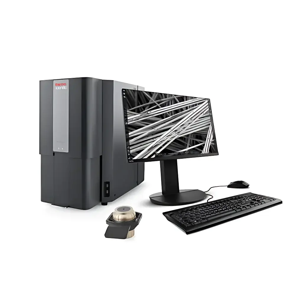
Phenom Pro Desktop Scanning Electron Microscope
| Brand | Phenom |
|---|---|
| Origin | Netherlands |
| Model | Phenom Pro |
| Instrument Type | Desktop SEM |
| Electron Source | CeB6 (Cerium Hexaboride) |
| Secondary Electron Resolution | 6 nm |
| Backscattered Electron Resolution | 6 nm |
| Magnification | Up to 350,000× |
| Accelerating Voltage | 4.8–20.5 kV |
| Vacuum Pump-down Time | <15 s |
| Filament Lifetime | >1,500 h |
| Imaging Modes | SE, BSE, and Real-time SE/BSE Mixed Imaging |
| Upgrade Path | Compatible with Phenom ProX EDS integration |
Overview
The Phenom Pro Desktop Scanning Electron Microscope is an engineered solution for laboratories requiring rapid, high-fidelity surface imaging without the infrastructure or operational overhead of floor-standing SEM systems. Based on thermionic emission from a cerium hexaboride (CeB6) electron source, the Phenom Pro operates under variable accelerating voltages (4.8–20.5 kV), enabling optimized contrast and penetration depth across diverse material classes—from insulating polymers to conductive metals and beam-sensitive biological specimens. Its compact vacuum architecture achieves pump-down in under 15 seconds, minimizing workflow interruption. Unlike conventional SEMs requiring dedicated rooms, vibration isolation tables, or high-voltage power conditioning, the Phenom Pro integrates all core electron-optical subsystems—including electrostatic lens column, dual-channel signal detection, and digital image acquisition—into a single benchtop enclosure. This design supports routine morphological characterization at resolutions ≤6 nm in both secondary electron (SE) and backscattered electron (BSE) modalities, meeting ISO 16700:2016 requirements for SEM resolution verification and ASTM E1558-22 guidelines for electron beam instrument qualification.
Key Features
- Real-time mixed-mode imaging: Simultaneous acquisition and display of SE and BSE signals, enhancing topographic and compositional contrast interpretation without manual mode switching.
- CeB6 thermionic source: Delivers stable beam current (>100 nA) with >1,500-hour operational lifetime, eliminating frequent filament replacement and reducing long-term cost of ownership.
- Integrated dual-detector system: High-efficiency Everhart-Thornley SE detector and solid-state BSE detector mounted within the chamber, enabling rapid contrast optimization and Z-contrast mapping.
- Automated workflow engine: One-click alignment, auto-focus, auto-stigmation, and stage calibration reduce operator dependency; no prior SEM experience required.
- Modular upgrade path: Hardware-ready for seamless integration of energy-dispersive X-ray spectroscopy (EDS) via the Phenom ProX conversion kit, supporting elemental mapping per ISO 14704:2022.
- Robust environmental tolerance: Operates reliably in standard lab environments (20–25°C, 30–70% RH), with no requirement for external cooling water or compressed air.
Sample Compatibility & Compliance
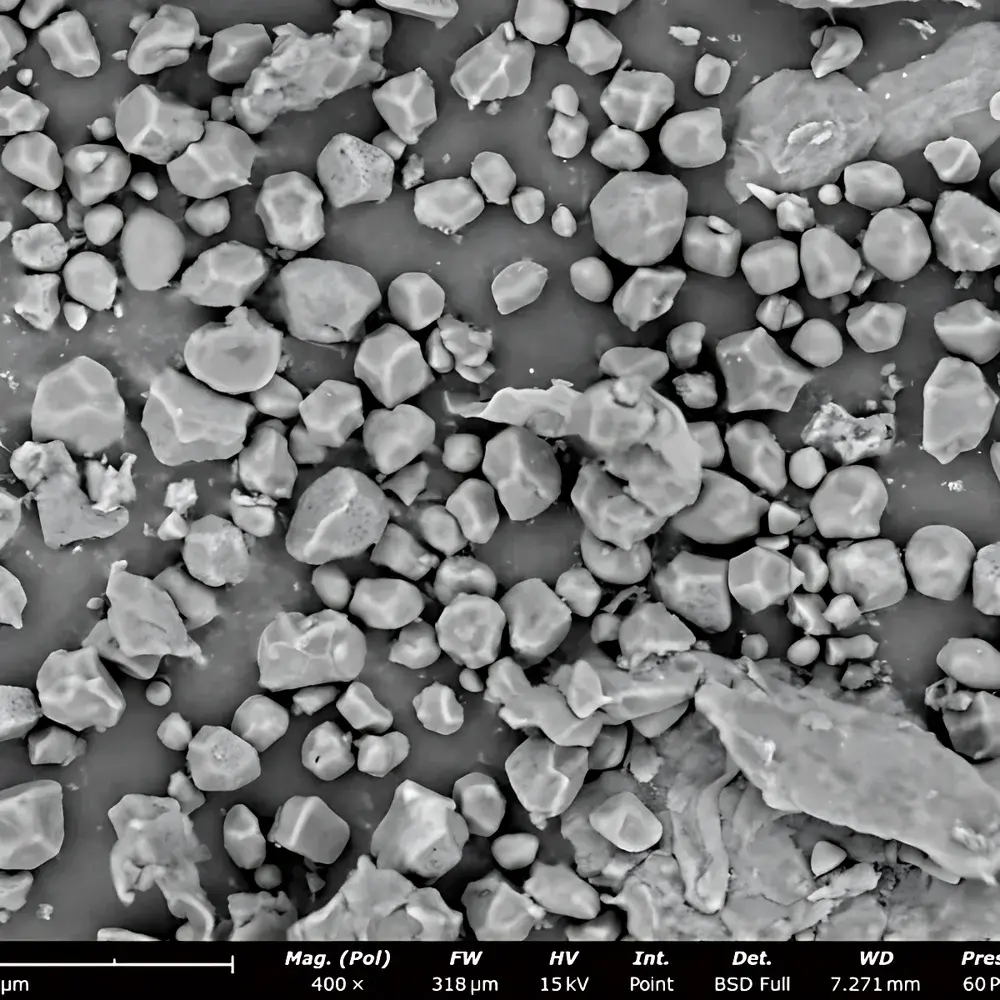
The Phenom Pro accommodates samples up to 100 mm in diameter and 50 mm in height, including non-conductive materials requiring minimal or no sputter coating (e.g., carbon or Au/Pd ≤5 nm). Its low-kV imaging capability (down to 4.8 kV) mitigates charging artifacts and preserves structural integrity of beam-sensitive specimens such as pharmaceutical powders, polymers, and hydrated tissues. All operational parameters—including dwell time, scan speed, and beam current—are fully adjustable to comply with GLP-compliant documentation workflows. The system meets IEC 61000-6-3:2019 (EMC emissions) and IEC 61000-6-2:2019 (immunity) standards. Data files are stored in vendor-neutral TIFF and CSV formats, supporting traceability under FDA 21 CFR Part 11 when paired with validated LIMS or ELN platforms.
Software & Data Management
Acquisition and analysis are managed through Phenom Desktop Software v5.x, a Windows-based application featuring intuitive ribbon-style navigation and scriptable measurement routines. Built-in tools include particle analysis (ISO 13322-1:2020 compliant), roughness quantification (Sa, Sq per ISO 25178-2), and automated report generation with embedded metadata (operator ID, timestamp, instrument serial number, acquisition parameters). Raw image data retains full 16-bit dynamic range and spatial calibration information. Export options include annotated PDF, Excel-compatible measurement logs, and 3D surface reconstruction exports compatible with third-party metrology software (e.g., MountainsMap®, Gwyddion). Audit trail functionality records all parameter changes and user actions, satisfying internal QA requirements for ISO/IEC 17025-accredited testing laboratories.
Applications
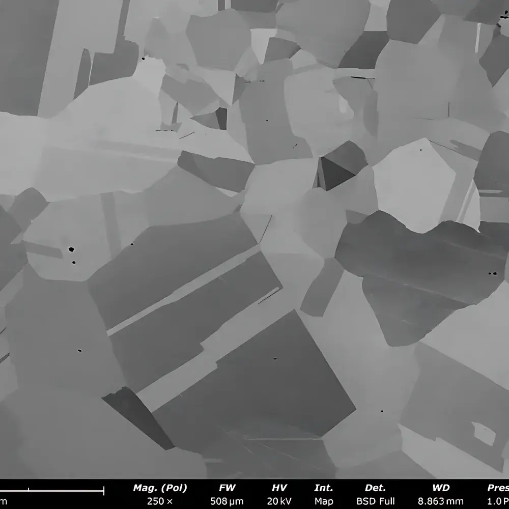
- Materials science: Fractography of alloys, grain boundary analysis in ceramics, dispersion uniformity assessment in nanocomposites.
- Electronics: Failure analysis of solder joints, contamination identification on PCB surfaces, cross-sectional imaging of thin-film stacks.
- Life sciences: Morphological evaluation of freeze-dried biologics, microbial colony topography, pollen grain ultrastructure.
- Geosciences: Mineral phase differentiation in sedimentary thin sections, pore network characterization in shale cores.
- Quality control: Incoming raw material inspection, coating thickness verification, wear debris analysis in lubricants.
FAQ
Is the Phenom Pro suitable for uncoated non-conductive samples?
Yes—low-kV imaging (4.8–10 kV) combined with charge compensation algorithms enables stable imaging of ceramics, polymers, and biological tissues without metallization in most cases.
Can the system be integrated into an existing laboratory information management system (LIMS)?
Yes—via configurable API endpoints and standardized file export protocols (TIFF, CSV, XML), supporting automated data ingestion and audit trail synchronization.
What vacuum level does the Phenom Pro achieve, and how is it maintained?
The system reaches a base pressure of ≤1 × 10⁻⁴ mbar using a turbomolecular pump backed by a dry scroll pump; vacuum integrity is continuously monitored and logged during operation.
Does the Phenom Pro support automated particle analysis for regulatory submissions?
Yes—the built-in particle module complies with ISO 13322-1:2020 and generates reports with statistical confidence intervals, measurement uncertainty estimates, and raw image archives required for FDA or EMA submissions.
How is software validation handled for GxP environments?
Phenom provides IQ/OQ documentation templates, installation qualification checklists, and a software validation guide aligned with ASTM E2500-22 and Annex 11 principles; site-specific PQ execution remains the responsibility of the end user.


