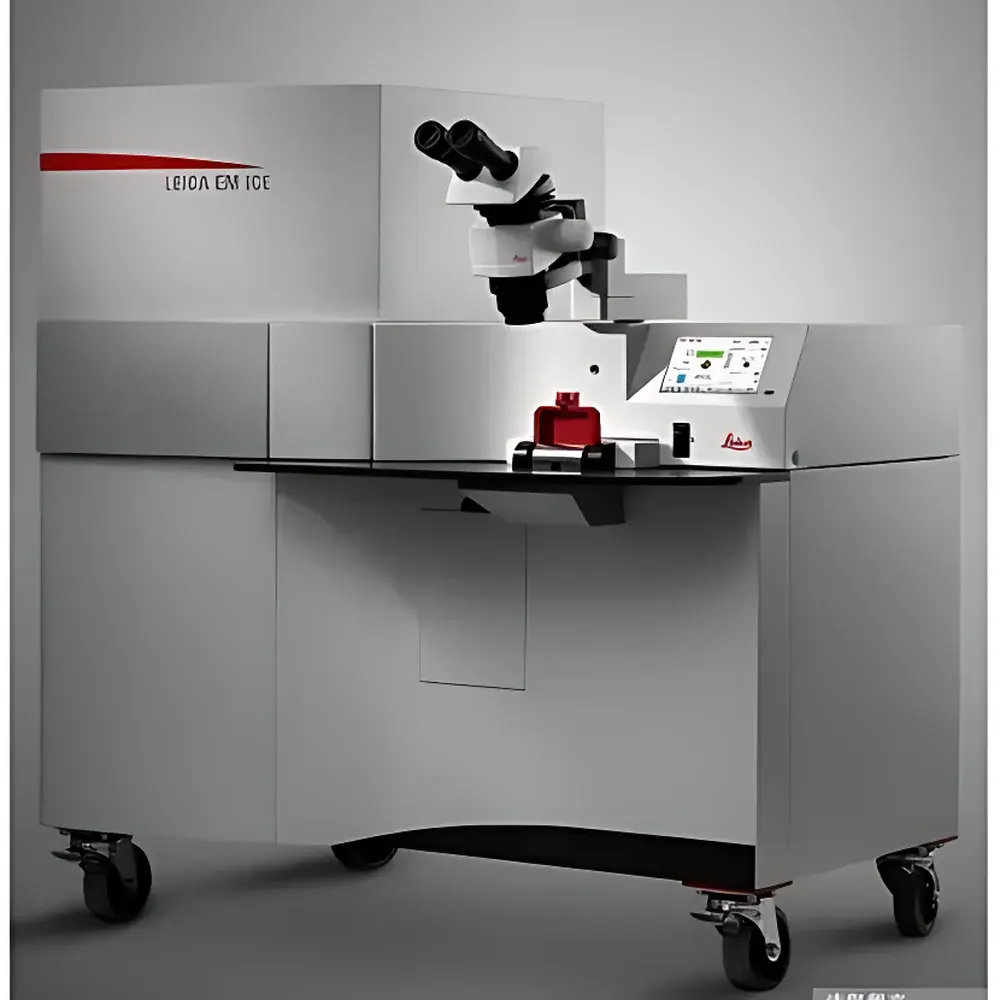
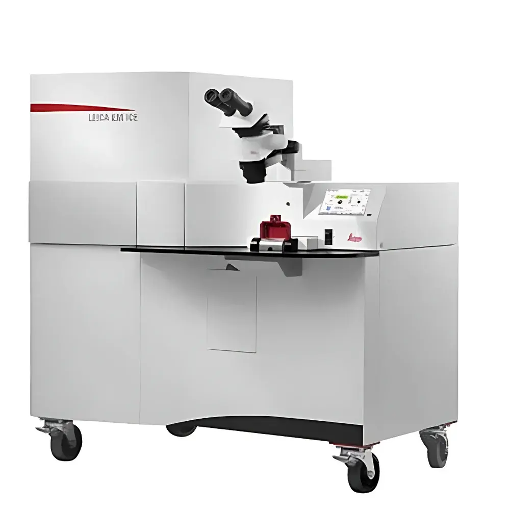
Leica EM ICE High-Pressure Freezer
| Brand | Leica |
|---|---|
| Origin | Germany |
| Model | Leica EM ICE |
| Type | Integrated Light-Stimulated High-Pressure Freezer |
| Cryogen Consumption (Daily, incl. cooling) | ≤30 L Liquid Nitrogen (LN₂) |
| Cooling Efficiency Improvement vs. Predecessor | ~70% reduction in LN₂ consumption during freezing |
| Pressure Range | Up to 210 MPa |
| Freeze-to-Stimulus Timing Precision | ≤5 ms |
| LED Wavelength Options | 380 nm (UV), 450 nm (Blue), 523 nm (Green), 597 nm (Amber), 660 nm (Red) |
| Sample Capacity | 9 specimens per session (3 positions × up to 3 specimens each) |
| Automation Level | Fully automated loading, alignment, pressurization, cryogen injection, and freezing sequence |
| Environmental Certification | DIN EN ISO 14001 certified |
| Packaging | Reusable, slope-integrated “Smart Packaging” (Austrian National Award for Intelligent Packaging, Nov. 2014) |
Overview
The Leica EM ICE is a fully integrated high-pressure freezer engineered for ultra-rapid vitrification of hydrated biological and soft-material specimens under physiologically relevant conditions. It operates on the principle of high-pressure freezing (HPF), where samples are subjected to pressures up to 210 MPa while simultaneously cooled by liquid nitrogen (LN₂), suppressing ice crystal formation and enabling near-native-state preservation at nanometer-scale structural fidelity. Unlike conventional plunge-freezing or cryo-fixation methods, HPF eliminates solute segregation and mechanical distortion, making it the gold-standard preparation technique for cryo-electron microscopy (cryo-EM), cryo-focused ion beam (cryo-FIB) milling, and correlative light and electron microscopy (CLEM). The EM ICE uniquely integrates synchronized optical stimulation—via five user-selectable, software-controlled LED modules—directly into the HPF workflow. This allows precise temporal coupling between photostimulation and freezing onset, with stimulus-to-freeze delays reproducibly controlled within ≤5 milliseconds. Such capability is indispensable for capturing transient molecular events—including synaptic vesicle fusion, photoactivated protein conformational changes, or light-triggered phase transitions in functional materials.
Key Features
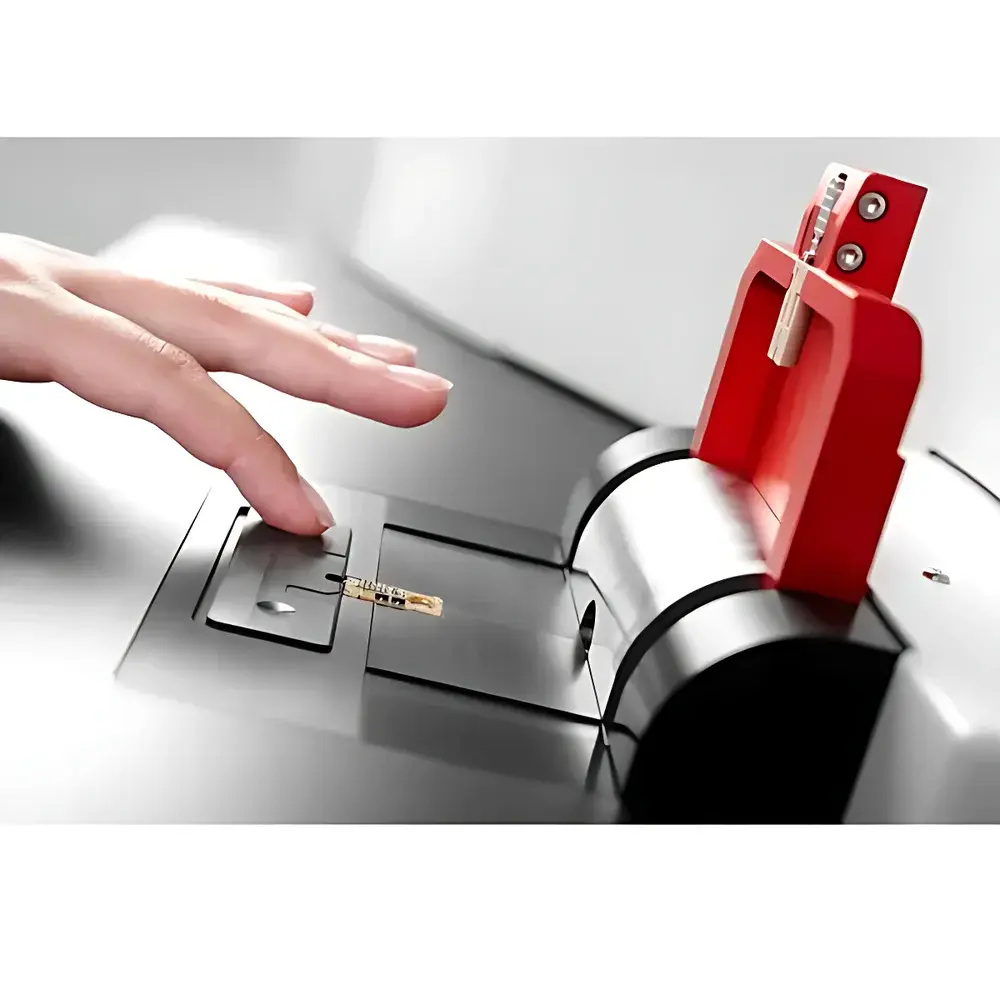
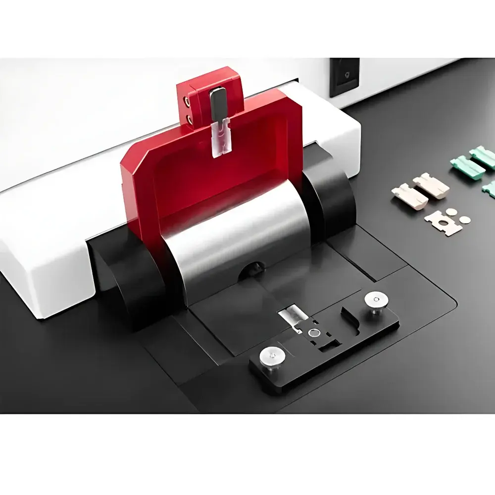
- Fully automated sample loading and alignment: Closure of the specimen stage lid initiates automatic cartridge engagement, pressure chamber sealing, and cryogen delivery—no manual intervention required.
- Integrated multi-wavelength LED stimulation system: Five interchangeable, auto-recognized LED modules (380 nm UV, 450 nm blue, 523 nm green, 597 nm amber, 660 nm red) enable wavelength-specific activation of optogenetic probes, photoswitches, or light-sensitive formulations (e.g., photopharmacological agents, photoresponsive polymers, or food-grade photosensitizers).
- Sub-millisecond temporal precision: Stimulus onset and freezing initiation are hardware-synchronized via TTL triggers; all timing parameters are logged with timestamped metadata in the acquisition log file.
- High-throughput capability: Three independent sample positions support up to nine consecutive freeze cycles without operator reconfiguration; each position permits programmable specimen count, rotation angle, and condition labeling.
- Low-consumption cryogenic architecture: Daily LN₂ consumption remains ≤30 L—including continuous system cooling—representing a ~65% reduction versus prior-generation HPF instruments; freezing-phase LN₂ usage is reduced by ~70% through optimized thermal management and pressure-cooling synergy.
- Modular upgrade path: Light-stimulation functionality and environmental control units (e.g., heated stage, climate-controlled chamber) can be installed on-site by Leica service engineers—no instrument return or downtime required.
Sample Compatibility & Compliance
The Leica EM ICE accommodates standard 3 mm diameter specimen carriers (e.g., aluminum planchettes, copper hats, or custom-designed holders) compatible with subsequent cryo-EM grid preparation, cryo-FIB sectioning, or freeze-fracture replication. It supports aqueous, semi-solid, and suspension-based samples—including cultured cells, tissue slices, organoids, bacterial biofilms, hydrogels, emulsions, and light-responsive industrial formulations (e.g., cosmetic actives, photo-curable resins, or food colloids). All operational protocols adhere to Good Laboratory Practice (GLP) principles. Audit-trail functionality—including user ID, timestamp, pressure profile, temperature trace, LED module identity, stimulation duration, and freeze delay—is automatically recorded and exportable for regulatory review. The system meets DIN EN ISO 14001 environmental management standards and complies with EU RoHS and WEEE directives. No alcohol or auxiliary cryogens are required—eliminating chemical handling risks and disposal liabilities.
Software & Data Management
Control and monitoring are executed via Leica’s proprietary EM ICE Control Software, a Windows-based application providing real-time visualization of pressure ramping, LN₂ level, chamber temperature, and LED status. Each freeze event generates a structured XML log containing full experimental metadata: operator name, date/time, specimen ID, position index, selected LED wavelength and intensity, stimulus duration, freeze delay, final pressure, and system health flags. Log files are digitally signed and time-stamped to support 21 CFR Part 11 compliance when deployed in regulated environments (e.g., pharmaceutical preclinical research or biologics characterization). Data export supports CSV, JSON, and HDF5 formats for integration with downstream analysis pipelines—including cryo-EM processing suites (RELION, cryoSPARC), CLEM correlation tools, or custom MATLAB/Python workflows.
Applications
- Cryo-EM structural biology: Capturing short-lived intermediates in enzymatic catalysis, GPCR activation, or ribosome assembly via light-triggered trapping.
- Neuroscience: Freezing synaptic terminals at defined millisecond latencies post-optogenetic stimulation to resolve vesicle pool dynamics and active zone architecture.
- Cell biology: Immobilizing cytoskeletal remodeling, organelle motility, or membrane trafficking events induced by caged compounds or channelrhodopsin activation.
- Materials science: Studying photoinduced phase separation, polymer crosslinking kinetics, or nanoparticle self-assembly in situ.
- Industrial R&D: Characterizing light-triggered texture changes in food matrices, photostability of cosmetic actives, or curing behavior of dental composites under native hydration.
FAQ
What is the minimum achievable stimulus-to-freeze delay?
The system achieves ≤5 ms temporal resolution between LED pulse onset and high-pressure freezing initiation, verified by embedded photodiode and piezoelectric pressure transducer synchronization.
Can I use non-Leica specimen carriers?
Yes—any carrier conforming to standard 3 mm outer diameter and flat-bottom geometry (e.g., Boekema-type, Reichelt-type, or custom-machined planchettes) is mechanically compatible.
Is the software compatible with LIMS or ELN systems?
Raw log files are exportable in open, schema-defined formats (CSV, JSON); API access for direct LIMS/ELN integration is available upon request through Leica’s Scientific Computing Group.
Does the EM ICE require external vibration isolation?
No—the instrument incorporates passive damping mounts and a rigid monocoque frame; operation on standard laboratory benches is sufficient when ambient vibrations remain below ISO 2631-2 Class A thresholds.
How is calibration maintained over time?
Pressure transducers and temperature sensors are factory-calibrated traceable to NIST standards; annual verification kits and on-site recalibration services are offered as part of Leica’s Extended Service Agreement.