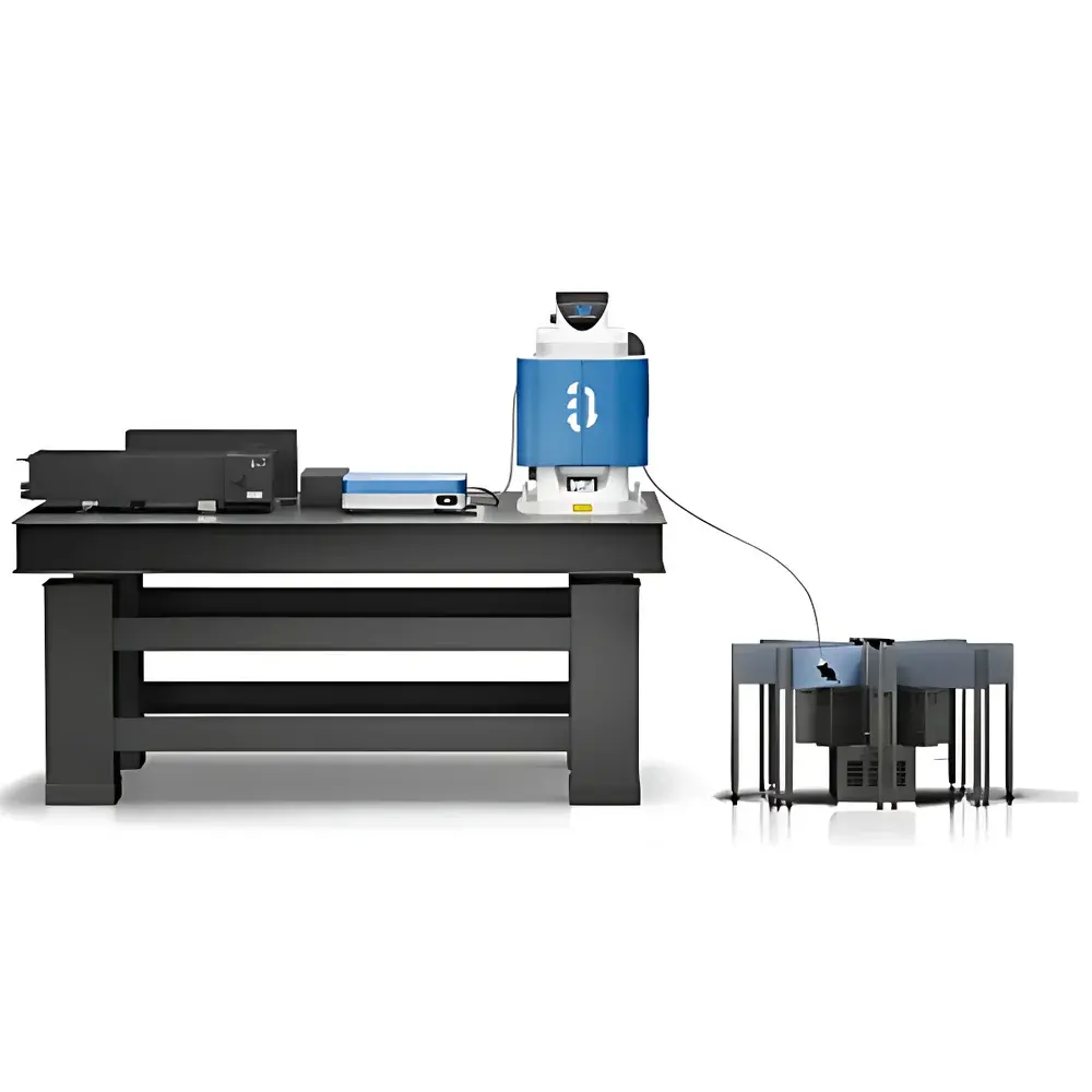
Auniontech SUPERNOVA-3000 Miniaturized Three-Photon Microscope
| Brand | Auniontech |
|---|---|
| Origin | Shanghai, China |
| Manufacturer Type | Authorized Distributor |
| Product Category | Domestic |
| Model | SUPERNOVA-3000 Biological In Vivo Imaging System |
| Form Factor | Miniaturized Three-Photon Probe |
| Probe Weight | 2.2 g |
| Excitation NA / Collection NA | 0.55 / 0.65 |
| Immersion Medium | Water or Silicone Oil |
| Field of View | 400 × 400 µm² |
| Working Distance | 1.75 mm |
| Probe Diameter | 3.4 mm |
| Fluorescence Detection | GaAsP PMT (300–720 nm) |
| Green Channel | 520 ± 25 nm |
| Red Channel | 625 ± 25 nm |
| Controller | ≥120 MS/s Sampling Rate |
| Fiber-Coupled AOM | <250 ns Response Time |
| XYZ Stage | Bidirectional Repeatability ≤1 µm |
| Excitation Wavelength Range | 490–550 nm |
| Widefield Camera | 1920 × 1200 px, 40 Hz Full-Frame |
| Laser Options | 1300 nm / 1700 nm Femtosecond Oscillators |
| Software Suite | SUPERGIN (Acquisition), SUPERANALY (Analysis) |
| System Footprint | 595 × 400 × 668 mm³ |
| Operating Environment | 20–30 °C, RH <60% |
Overview
The Auniontech SUPERNOVA-3000 is a purpose-engineered miniaturized three-photon microscope designed for high-fidelity, deep-tissue functional imaging in freely behaving rodents. Unlike conventional two-photon systems limited by scattering-induced signal attenuation beyond ~600 µm, the SUPERNOVA-3000 leverages third-order nonlinear excitation at extended near-infrared wavelengths (e.g., 1300 nm and 1700 nm) to achieve unprecedented penetration depth—up to 1.2 mm in mouse brain tissue—enabling direct calcium imaging of hippocampal CA1 subfields through intact cortex and corpus callosum. Its optical architecture integrates an on-probe Abbe condenser collinear with a simplified infinity-corrected objective, significantly enhancing collection efficiency of multiply scattered fluorescence photons from deep layers. This design circumvents the fundamental trade-off between resolution, working distance, and photon collection in miniaturized in vivo platforms, while maintaining compatibility with standard cranial window preparations—eliminating the need for invasive GRIN lens implantation.
Key Features
- Deep-tissue access: Achieves 1.2 mm imaging depth in mouse brain using 1300/1700 nm femtosecond excitation—sufficient to span cortex, corpus callosum, and hippocampal CA1 without tissue resection.
- Ultra-lightweight probe: 2.2 g total mass enables stable head-mounting on adult mice during naturalistic behaviors including locomotion, whisking, and skilled forelimb tasks.
- Scattering-optimized optics: Integrated Abbe condenser and shared-tube-lens configuration increase scattered photon throughput by >2× versus conventional miniaturized two-photon probes.
- True 3D volumetric imaging: Electrotunable lens (ETL)-based axial scanning enables rapid z-stack acquisition without mechanical stage movement—critical for motion-robust in vivo operation.
- Low phototoxicity operation: Longer excitation wavelengths reduce linear absorption and multiphoton-induced photodamage; enhanced collection efficiency permits lower incident power (e.g., 35.9 mW at objective back aperture for 978 µm CA1 imaging).
- Modular laser integration: Supports OEM coupling to commercial femtosecond oscillators via air-core photonic crystal fiber and fast AOM gating (<250 ns rise time), ensuring pulse fidelity and laser safety compliance.
Sample Compatibility & Compliance
The SUPERNOVA-3000 is validated for longitudinal in vivo imaging in transgenic and virally labeled mice expressing genetically encoded calcium indicators (e.g., GCaMP6s, RCaMP, jRGECO1a) or structural labels (e.g., tdTomato, mCherry). It operates under standard cranial window protocols (thinned-skull or glass-embedded), eliminating requirements for chronic GRIN lens implantation and associated glial scarring or neuronal displacement. The system complies with ISO 13485-aligned manufacturing controls for research instrumentation and meets IEC 60825-1:2014 Class 4 laser safety requirements when operated with interlocked shutters and beam path containment. Data acquisition workflows support GLP-compliant metadata tagging (timestamp, laser power, Z-position, behavioral trigger sync) and are compatible with FDA 21 CFR Part 11 audit trail configurations when deployed with validated SUPERGIN/SUPERANALY software versions.
Software & Data Management
The SUPERNOVA-3000 is controlled by SUPERGIN—a real-time acquisition platform built on LabVIEW RT and FPGA-based timing cores, delivering deterministic synchronization between ETL voltage ramps, galvanometric scanning, PMT digitization (≥120 MS/s, 14-bit), and external behavioral triggers (TTL, analog, USB). Acquired data are stored in HDF5 format with embedded provenance metadata (laser parameters, objective position, environmental logs). SUPERANALY provides modular processing pipelines for motion correction (non-rigid registration), ROI segmentation (CNMF-E), deconvolution-based spike inference, and cross-modal alignment with video tracking (DeepLabCut integration). Both applications support batch processing on Windows 10 workstations (32 GB RAM, dual-drive storage: 512 GB NVMe + 2 TB HDD) and export standardized NWB 2.0 files for interoperability with Neurodata Without Borders ecosystem tools.
Applications
- Multi-layer cortical dynamics: Simultaneous recording across L1–L6 in parietal and visual cortices during sensory stimulation or decision-making tasks.
- Hippocampal–cortical circuit mapping: Direct observation of CA1 neuron activity correlated with prefrontal or entorhinal inputs during spatial navigation and memory recall.
- Skilled motor behavior: High-speed (≥15 Hz) volumetric imaging of layer VI neurons in posterior parietal cortex during sugar pellet retrieval through narrow apertures.
- Neurovascular coupling: Concurrent detection of GCaMP6s calcium transients and third-harmonic generation (THG) signals from dura, microvasculature, and white matter interfaces—enabling intrinsic structural referencing without exogenous dyes.
- Longitudinal plasticity studies: Repeated imaging over weeks through same cranial window to track dendritic spine turnover, axonal bouton dynamics, or network reorganization post-injury or learning.
FAQ
What is the maximum achievable imaging depth in mouse brain tissue?
The SUPERNOVA-3000 achieves consistent functional calcium imaging at depths up to 1.2 mm below the pial surface—verified in vivo across multiple animals using 1700 nm excitation and GCaMP6s expression in hippocampal CA1.
Does the system require GRIN lens implantation?
No. The probe is designed for use with standard cranial window preparations only—avoiding surgical complications associated with GRIN lens insertion and enabling faster recovery and higher experimental throughput.
Can the probe be used with existing femtosecond laser sources?
Yes. The integrated fiber-coupling module accepts free-space or fiber-pigtailed oscillators (e.g., Spectra-Physics Mai Tai, Coherent Chameleon, or Toptica FemtoFiber) via customizable collimation and AOM triggering interfaces.
How is motion artifact corrected during freely moving experiments?
Real-time motion correction is implemented via hardware-synchronized ETL z-scanning and post-hoc non-rigid image registration in SUPERANALY, leveraging both fluorescence and THG structural landmarks for sub-micron alignment accuracy.
Is the software compliant with regulatory data integrity standards?
SUPERGIN and SUPERANALY support configurable audit trails, electronic signatures, and data immutability settings aligned with 21 CFR Part 11 requirements for regulated preclinical studies.


