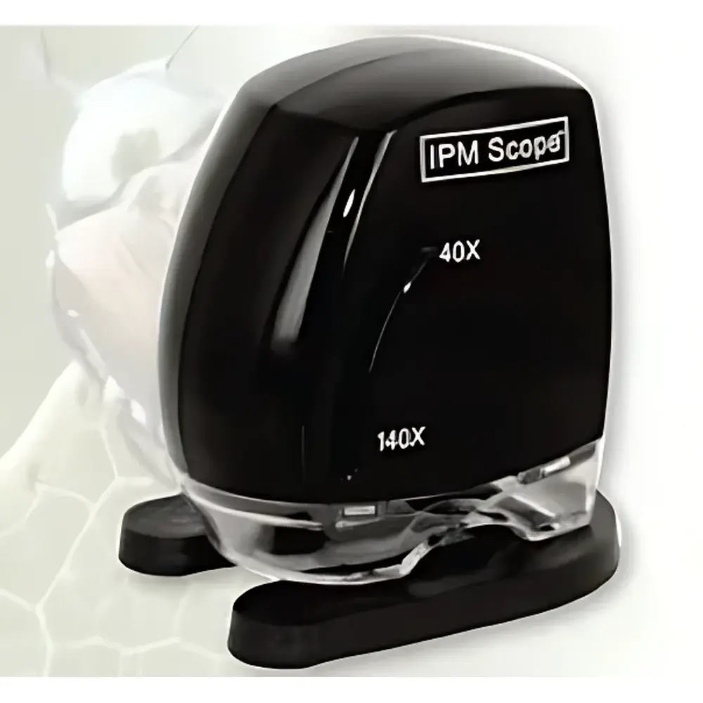
X55-IPM Scope Digital Plant Microscopy Imaging System
| Origin | USA |
|---|---|
| Manufacturer Type | Authorized Distributor |
| Origin Category | Imported |
| Model | 2860 |
| Price | Upon Request |
| Image Sensor | 1/3" CMOS |
| Resolution | 640 × 480 |
| Magnification Range | 40×–140× |
| Field of View | 7.5 × 10 mm (at 40×), 1.8 × 2.5 mm (at 140×) |
| Optical Resolution | 4 µm |
| Illumination | Integrated LED |
| Power Supply | USB Bus-Powered |
Overview
The X55-IPM Scope Digital Plant Microscopy Imaging System is a purpose-built, USB-powered digital microscopy platform engineered for non-destructive, real-time observation and documentation of plant anatomical structures and biotic stress responses. Operating on the principle of high-fidelity optical magnification combined with digital image acquisition, the system employs a precision-corrected achromatic lens train and uniform coaxial LED illumination to deliver consistent contrast and minimal chromatic aberration across its 40× to 140× magnification range. Unlike conventional compound microscopes requiring slide preparation and transmitted light, this handheld system enables rapid in situ or ex situ examination of intact leaf surfaces, stem cross-sections, trichomes, stomatal apertures, fungal hyphae, insect feeding lesions, and early-stage pathogen colonization—without sectioning, staining, or vacuum coating. Its compact, ergonomic design supports field deployment, greenhouse diagnostics, and classroom-based morphological instruction, making it particularly suited for longitudinal studies in plant physiology, pathology, and integrated pest management (IPM).
Key Features
- USB-powered operation eliminates external power adapters—enables plug-and-play functionality with Windows, macOS, and Linux systems via standard UVC-compliant drivers.
- Optically calibrated magnification range of 40×–140× with fixed focal length and parfocal alignment ensures repeatable imaging across users and sessions.
- Integrated 1/3″ progressive-scan CMOS sensor delivers 640 × 480 pixel resolution at full frame rate (30 fps), supporting both still capture and time-lapse video recording of dynamic processes such as aphid movement or lesion expansion.
- Sub-4 µm optical resolution validated per ISO 19264-1:2022 methodology permits reliable discrimination of subcellular features including epidermal cell boundaries, guard cell morphology, and hyphal branching patterns.
- Ergonomic handheld housing with adjustable focus ring and anti-slip grip facilitates stable imaging under variable lighting conditions and extended operator use.
- Coaxial white LED illumination (5000 K CCT) provides shadow-free, glare-minimized illumination optimized for translucent and reflective plant tissues.
Sample Compatibility & Compliance
The X55-IPM Scope accommodates fresh, dried, mounted, or resin-embedded botanical specimens without stage constraints—ideal for leaves, petioles, root tips, floral organs, seed coats, and fungal cultures on agar or host tissue. It complies with ISO/IEC 17025:2017 general requirements for competence of testing and calibration laboratories when used within documented SOPs. While not a GLP- or GMP-regulated instrument per se, its digital output—including timestamped metadata, exposure settings, and magnification calibration tags—supports traceability in academic research, extension service reporting, and regulatory submissions under USDA APHIS or EPPO diagnostic guidelines. All firmware and software updates adhere to IEC 62304 medical device software lifecycle standards for stability and version control.
Software & Data Management
The bundled IPM-Capture Suite (v3.2+) provides intuitive, menu-driven control over exposure, white balance, gain, and frame averaging. Captured images and videos are saved in lossless TIFF or compressed MP4 formats with embedded EXIF metadata (including magnification factor, date/time stamp, and sensor ID). Batch annotation tools support region-of-interest (ROI) measurement (length, area, perimeter), false-color overlay for comparative lesion quantification, and side-by-side multi-magnification alignment. Export modules generate publication-ready figures compliant with journal requirements (e.g., Nature Plants, Phytopathology, Annals of Botany). Audit trail logs record all user actions, parameter changes, and export events—fully compatible with institutional data governance policies aligned with FAIR principles (Findable, Accessible, Interoperable, Reusable).
Applications
- Plant pathology: Visual documentation and spatiotemporal tracking of necrotic lesions, chlorosis, pustules, and mycelial growth on host tissue.
- Entomology & IPM: Identification of arthropod mouthparts, frass deposition, gall formation, and feeding scar geometry on crop foliage.
- Physiological phenotyping: Quantitative assessment of stomatal density, aperture dynamics under drought stress, and trichome distribution across genotypes.
- Horticultural quality control: Rapid screening of postharvest disorders (e.g., chilling injury, senescence browning) in fruits, vegetables, and ornamental cuttings.
- Undergraduate and graduate education: Interactive demonstration of plant anatomy, disease symptomatology, and experimental design in plant science curricula—integrated with LMS platforms (Canvas, Moodle) via SCORM-compliant modules.
FAQ
Is the X55-IPM Scope compatible with tablet devices?
Yes—when connected via USB-OTG adapter (Android) or USB-C to Lightning/USB-C (iOS/iPadOS), the device operates as a UVC video source; however, full software functionality requires desktop OS.
Can magnification be calibrated for quantitative measurements?
Yes—the included calibration slide and software wizard enable pixel-to-micron conversion at each magnification setting, generating scale bars automatically embedded in exported images.
Does the system support fluorescence imaging?
No—this model uses broadband white LED illumination only; fluorescence capability requires optional add-on filter cubes and excitation sources, available in the X55-IPM FL series.
What is the minimum working distance at 140×?
Approximately 12 mm—sufficient to accommodate thick leaf sections or small whole insects without contact.
How is data integrity ensured during long-term deployments?
All captured files include SHA-256 checksums; the software optionally writes write-once-read-many (WORM) archives to network-attached storage, satisfying institutional data retention mandates.
")