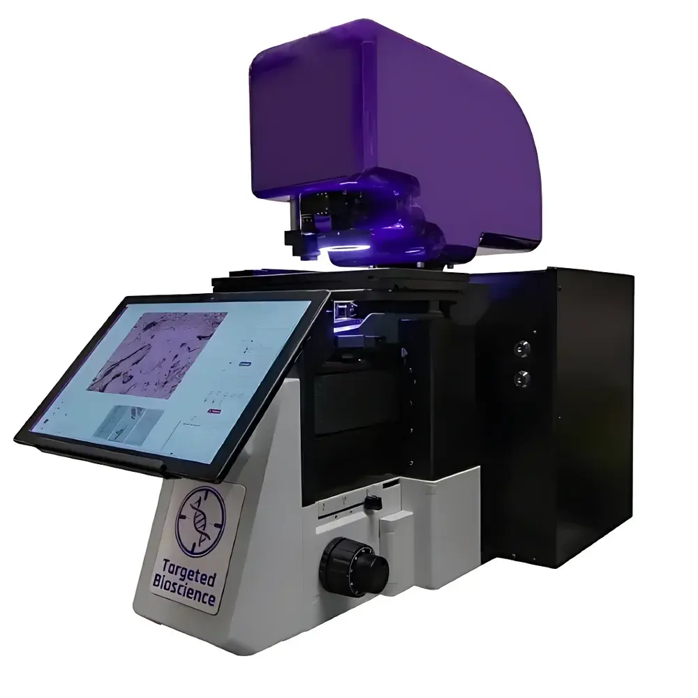
AccuLift™ Laser Microdissection and Capture System
| Brand | — |
|---|---|
| Origin | USA |
| Manufacturer Type | Authorized Distributor |
| Origin Category | Imported |
| Model | AccuLift |
| Pricing | Upon Request |
| Separation Method | Automated |
| Microscope Platform | Olympus IX73 or Nikon Ti2 base |
| IR Capture Laser | 808 nm solid-state, adjustable power |
| UV Cutting Laser | 355 nm & 349 nm diode-pumped Q-switched solid-state, tunable current & pulse frequency |
| Illumination | 5W 24-LED ring light |
| Objectives | Olympus PLAPON/PLANAPO 2×, 4×, 10×, 20×, 40×, 60×, 100× (dry) |
| Stage | Motorized X/Y stage with 0.25 µm homing accuracy & bidirectional repeatability |
| Camera | Dual 5 MP CMOS (color & monochrome), 75 fps at full resolution, large FOV, low-noise |
| Contrast Mode | Brightfield |
| Software Interface | Proprietary LCM control suite with intuitive GUI |
Overview
The AccuLift™ Laser Microdissection and Capture System is a dual-wavelength, research-grade instrument engineered for precise spatial isolation of morphologically defined cells or tissue regions under direct microscopic visualization. It integrates infrared (IR) laser-based thermoplastic capture and ultraviolet (UV) laser ablation in a single platform—enabling both non-contact transfer of intact cells and high-fidelity microsurgical excision. The system operates on the principle of selective photothermal activation: the 808 nm IR laser induces localized melting of a thermolabile polymer film coated on specialized cap or slide substrates, allowing adhesion of targeted cells without mechanical contact or thermal damage to biomolecular integrity. Concurrently, the 355 nm and 349 nm UV lasers deliver focused, short-pulse energy for clean, sub-micron precision cutting—minimizing collateral thermal diffusion and preserving nucleic acid and protein quality for downstream multi-omics applications.
Key Features
- Dual-laser architecture: Independent, co-aligned 808 nm IR capture and 355/349 nm UV cutting lasers with real-time power and pulse parameter control.
- Flexible substrate compatibility: Supports proprietary polymer-coated collection caps, metal-frame membrane slides, standard glass microscope slides—reducing consumables cost without compromising capture efficiency.
- Motorized high-precision stage: X/Y travel with 0.25 µm homing accuracy and bidirectional repeatability; integrated “click-to-cut” navigation for rapid region-of-interest (ROI) selection.
- Optimized optical path: Compatible with Olympus IX73 or Nikon Ti2 inverted microscope bases; equipped with brightfield illumination (5 W 24-LED ring), and a full suite of dry objectives (2×–100×) for scalable magnification and depth-of-field control.
- Dual high-speed imaging: Simultaneous color and monochrome 5 MP CMOS cameras (75 fps at full resolution), enabling real-time morphological assessment and contrast-optimized targeting in heterogeneous tissue sections.
- Intuitive software interface: Proprietary LCM control software with drag-and-drop ROI annotation, batch processing, audit-trail logging, and export-compatible metadata tagging aligned with FAIR data principles.
Sample Compatibility & Compliance
The AccuLift system accommodates routinely processed FFPE and frozen tissue sections (4–20 µm thickness), cytospin preparations, and cultured monolayers. Its gentle IR capture mechanism preserves RNA integrity (RIN > 8.5) and native protein conformation—validated per ISO 20387:2018 (biobanking) and compatible with workflows adhering to CLIA, CAP, and GLP guidelines. All consumables—including polymer-coated caps and extraction kits—are manufactured under ISO 13485-certified conditions and supplied with CoA documentation. The software supports 21 CFR Part 11-compliant user authentication, electronic signatures, and immutable audit trails for regulated environments requiring traceability in clinical or translational research settings.
Software & Data Management
The AccuLift Control Suite provides an integrated environment for acquisition, annotation, execution, and export. Users define ROIs via freehand drawing, polygonal selection, or auto-threshold segmentation based on pixel intensity. Each dissection event is timestamped and linked to objective magnification, laser parameters, stage coordinates, and operator ID. Export formats include TIFF (annotated images), CSV (spatial coordinate logs), and JSON (structured metadata), ensuring interoperability with downstream bioinformatics pipelines (e.g., Seurat, CellxGene, MaxQuant). Raw image and parameter logs are stored in hierarchical folder structures compliant with BIDS and MIAME standards, facilitating institutional data repository ingestion and long-term archival.
Applications
- Tumor Microenvironment Mapping: Isolation of tumor-infiltrating lymphocytes (TILs), cancer-associated fibroblasts (CAFs), and malignant epithelial nests from multiplex IHC-stained sections for parallel WGS, RNA-seq, and TCR repertoire analysis.
- Neuroanatomical Profiling: Targeted retrieval of layer-specific neurons, glia, or amyloid plaque-associated microglia from human postmortem brain sections for spatial proteomics (LC-MS/MS) and single-cell epigenomics.
- Developmental Biology: Microdissection of embryonic germ layers or organ primordia from cryosections for time-resolved transcriptomic trajectories.
- Pathogen-Host Interactions: Spatial isolation of infected vs. bystander cells in viral or bacterial infection models to resolve cell-type-specific host response signatures.
- Clinical Biomarker Discovery: Retrieval of histopathologically validated lesion subregions (e.g., ductal carcinoma in situ vs. invasive front) for validation-grade qPCR or digital spatial profiling (RPPA).
FAQ
What types of tissue sections are compatible with the AccuLift system?
FFPE and OCT-embedded frozen sections (4–20 µm), cytology smears, and adherent cell cultures on standard glass slides or membrane-coated substrates.
Can the system be integrated with existing microscope platforms?
Yes—it is fully compatible with Olympus IX73 and Nikon Ti2 inverted microscope bases; custom mounting adapters are available for other OEM systems upon request.
Does the software support automated batch processing?
Yes—users can queue multiple slides with predefined ROI templates, laser settings, and stage coordinates for unattended overnight operation.
How is biomolecular integrity verified post-capture?
System validation includes RT-qPCR amplification of long amplicons (>500 bp), Bioanalyzer RNA integrity assays, and SDS-PAGE/Western blotting for protein yield and degradation markers—data included in the Installation Qualification (IQ) report.
Is remote operation or telepathology support available?
The software supports secure VNC-based remote access for collaborative annotation and supervision; however, laser execution requires local physical presence per laser safety Class 1M compliance requirements.


