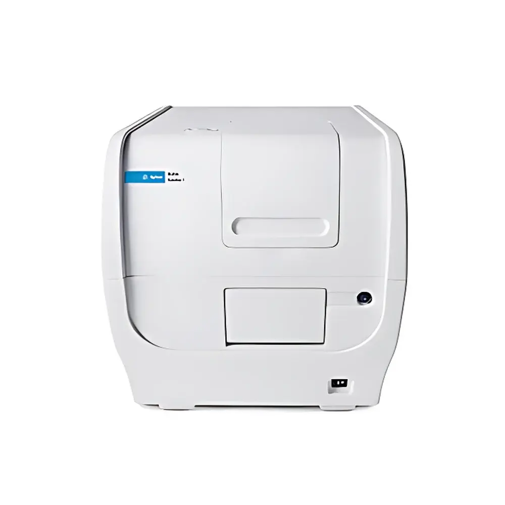
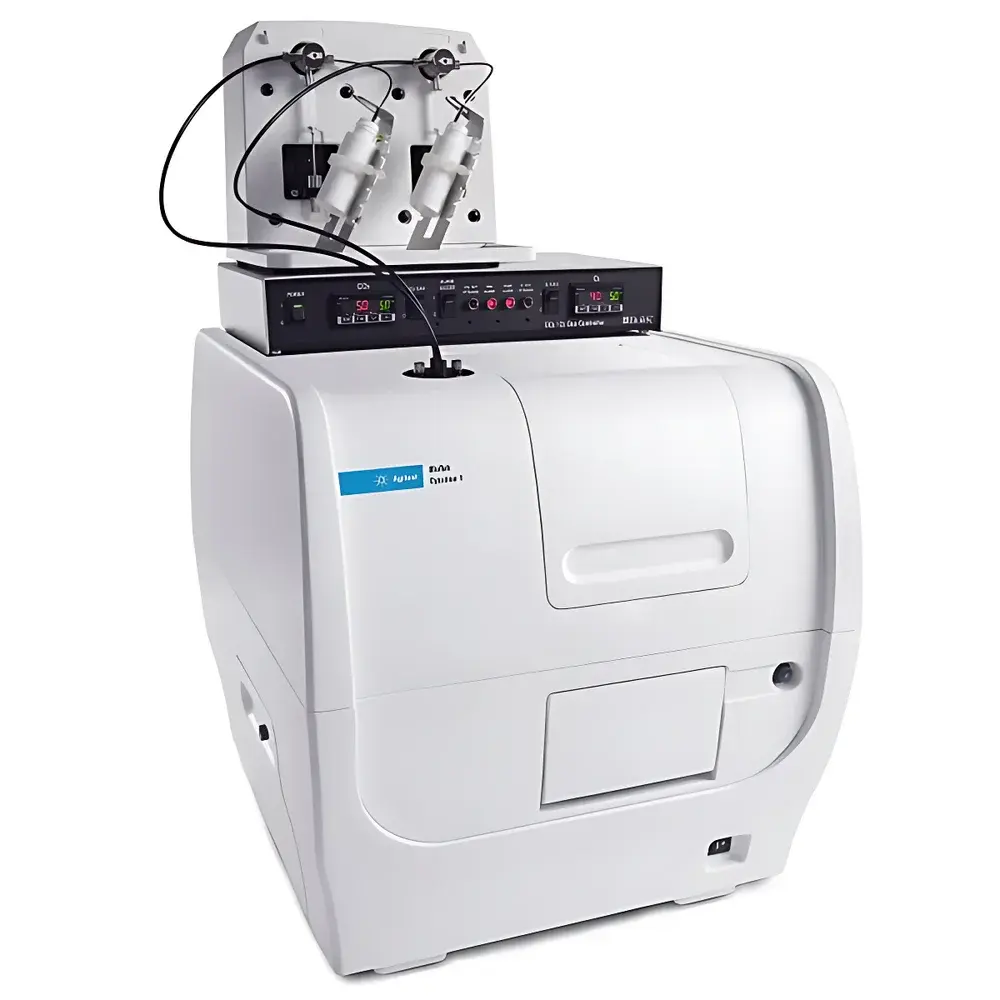
Agilent BioTek Cytation 1 Cell Imaging Multi-Mode Reader
| Brand | Agilent Technologies |
|---|---|
| Origin | USA |
| Manufacturer Type | Original Equipment Manufacturer (OEM) |
| Product Origin | Imported |
| Model | Cytation 1 |
| Temperature Control | Four-zone incubation up to 45 °C |
| Humidity Control | Integrated with four-zone incubation system |
Overview
The Agilent BioTek Cytation 1 Cell Imaging Multi-Mode Reader is an integrated, benchtop platform engineered for quantitative high-content cellular analysis in microplate format. It combines automated digital microscopy—supporting fluorescence and high-contrast brightfield imaging—with conventional multi-mode detection technologies including filter-based fluorescence intensity (FI), luminescence (LUM), and monochromator-based UV-Vis absorbance (ABS) measurements. Its core architecture implements a hybrid optical design: a fixed-objective, motorized XY stage with laser-assisted autofocus enables precise z-stack acquisition across 6- to 1536-well plates, while the dual-path detection system permits simultaneous or sequential acquisition of phenotypic image data and plate-based quantitative signals. This convergence supports kinetic, endpoint, and multiplexed assays under physiologically relevant conditions—making it suitable for applications ranging from cell viability and proliferation to subcellular translocation and morphometric profiling.
Key Features
- Integrated imaging and detection: Simultaneous acquisition of high-resolution fluorescence/brightfield images and quantitative microplate readouts (FI, LUM, ABS) within a single instrument workflow.
- Laser-based autofocus: Proprietary optical autofocus system utilizing reflected laser light from the liquid–air interface; delivers rapid, reproducible focusing independent of fluorescent signal intensity—critical for low-expression targets and long-term live-cell imaging.
- Four-zone environmental control: Independent temperature regulation (ambient to 45 °C), Peltier-driven cooling, condensation suppression, and optional CO₂/O₂ gas mixing modules enable stable, compartmentalized incubation for heterogeneous assay conditions across a single plate.
- Flexible optical configuration: Interchangeable LED/filter cube modules (over 20 configurations available); monochromator-based absorbance detection with 1 nm wavelength resolution (200–999 nm); programmable LED intensity and exposure time per well.
- Automated stage and acquisition: Motorized XY stage with sub-micron positioning repeatability; auto-exposure, auto-LED intensity adjustment, and batch image capture protocols reduce operator dependency and improve inter-run consistency.
Sample Compatibility & Compliance
The Cytation 1 accommodates standard microplate formats (6–1536-well), including clear-bottom, black, white, and specialized tissue-culture treated plates. It supports adherent and suspension cultures, spheroids, and organoid models when used with compatible plate types and environmental controls. The system complies with Good Laboratory Practice (GLP) and Good Manufacturing Practice (GMP) documentation requirements when operated with Gen5 software audit trail and electronic signature modules enabled. Data integrity aligns with FDA 21 CFR Part 11 expectations for secure user access, change tracking, and immutable result archiving. All thermal and gas control subsystems are calibrated and validated per Agilent’s ISO 9001-certified quality management system.
Software & Data Management
Gen5 Microplate Reader and Imaging Software serves as the unified control and analysis environment. It provides instrument control, protocol scripting, real-time image preview, and integrated analysis modules—including confluence measurement, object segmentation, intensity quantification, and morphology parameter extraction (e.g., nuclear area, cytoplasmic texture, neurite length). Batch processing supports multi-plate alignment, normalization, and export to CSV, TIFF, or OME-TIFF formats compliant with Bio-Formats. Advanced features include time-lapse macro scripting, ROI-based kinetic plotting, and direct integration with third-party analysis platforms such as ImageJ/Fiji and MATLAB via API. Audit trail logging records all user actions, parameter changes, and result modifications with timestamps and operator IDs.
Applications
The Cytation 1 is routinely deployed in academic, pharmaceutical, and biotechnology laboratories for: live-cell kinetic assays (e.g., calcium flux, mitochondrial membrane potential, apoptosis progression); label-free and stained cell counting and confluence monitoring; high-content screening of siRNA/CRISPR libraries; immunofluorescence-based protein localization; wound-healing and migration assays; and toxicity profiling using multiplexed endpoints (e.g., ATP content + nuclear morphology + ROS generation). Its ability to maintain physiological relevance during extended acquisitions—combined with hardware-level synchronization between imaging and detection events—ensures high reproducibility across biological replicates and experimental batches.
FAQ
Does the Cytation 1 support confocal imaging?
No—the Cytation 1 employs widefield fluorescence and brightfield optics. Confocal capability is available in higher-tier platforms such as the Cytation 5 and Lionheart FX.
Can Gen5 software perform machine learning-based image analysis?
Gen5 includes rule-based segmentation and feature extraction; for deep learning workflows (e.g., U-Net segmentation), users export annotated TIFF stacks to external frameworks like CellProfiler or Python-based tools.
Is CO₂ control included by default?
CO₂ and O₂ regulation are optional add-on modules—standard configurations include only temperature and humidity control via the four-zone Peltier system.
What is the maximum imaging magnification supported?
The system supports objectives from 2× to 60× (dry), with native pixel resolution down to ~0.6 µm/pixel at 60× magnification using the 5.0 MP scientific CMOS camera.
How does laser autofocus differ from contrast-based autofocus?
Laser autofocus measures physical distance to the meniscus—not sample contrast—enabling reliable focusing even in dim or non-fluorescent samples, and eliminating focus drift caused by photobleaching or media evaporation over time.
Related Products


