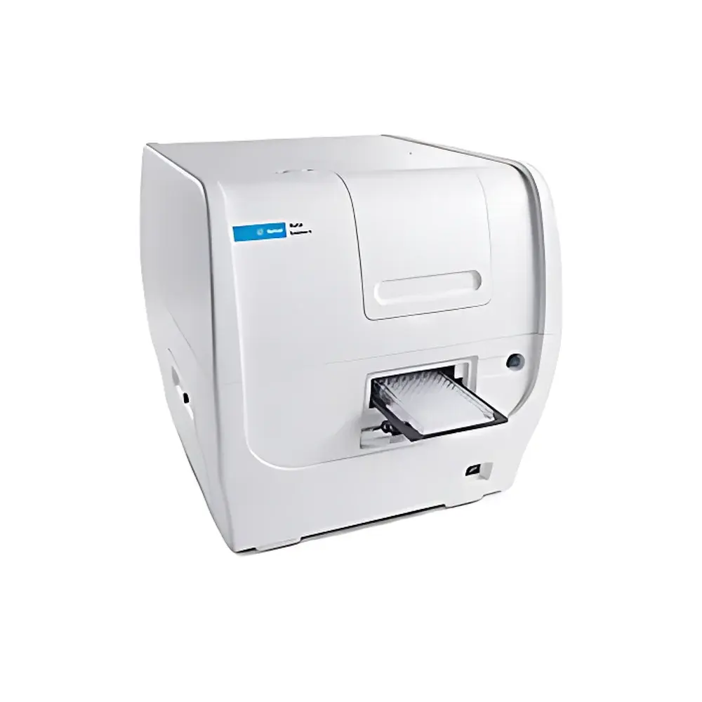
Agilent BioTek Cytation 5 Cell Imaging Multi-Mode Reader
| Brand | Agilent Technologies |
|---|---|
| Origin | USA |
| Manufacturer Type | Original Equipment Manufacturer (OEM) |
| Product Category | Imported Instrument |
| Model | Cytation 5 |
| Temperature Control Range | Up to 45 °C (Incubation) |
| Imaging Magnification | 1.25× to 60× (Air Objectives), 4× to 40× (Phase Contrast) |
| Microscope Type | Inverted |
| Camera | Sony CMOS, 16-bit color |
| Supported Vessels | 6–1536-well plates, slides, Petri dishes, T25 flasks, hemocytometers, chambered coverslips, Take3/Take3 Trio microplates |
| Environmental Control | Quad-zone temperature control (up to 65 °C), condensation management, CO₂/O₂ regulation, Peltier cooling |
| Detection Modes | UV-Vis absorbance, fluorescence intensity (filter & monochromator), luminescence, time-resolved fluorescence (TRF), TR-FRET, AlphaScreen/AlphaLISA, BRET, fluorescence polarization (FP) |
| Monochromator | Quad-grating system with tunable bandwidth |
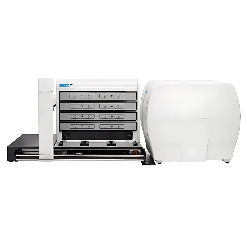
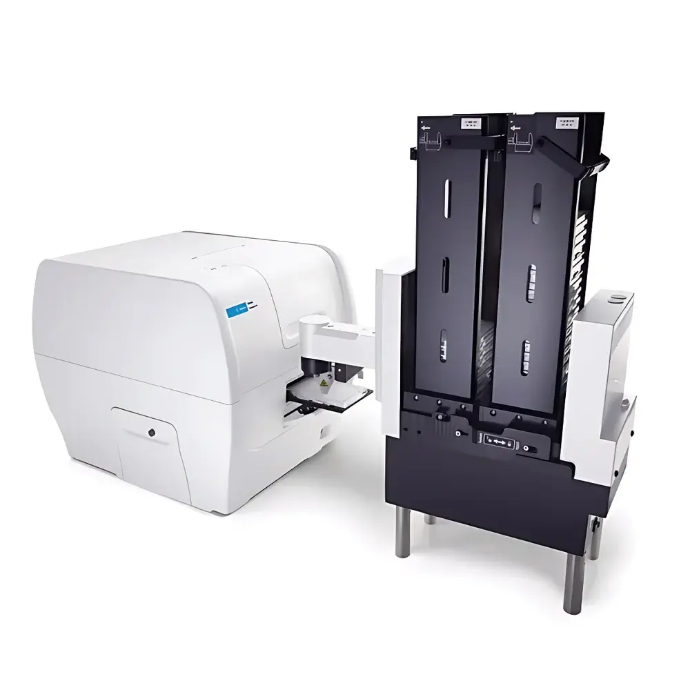
| Automation Compatibility | BioSpa 8, BioStack, BenchCel, third-party robotic handlers |
Overview
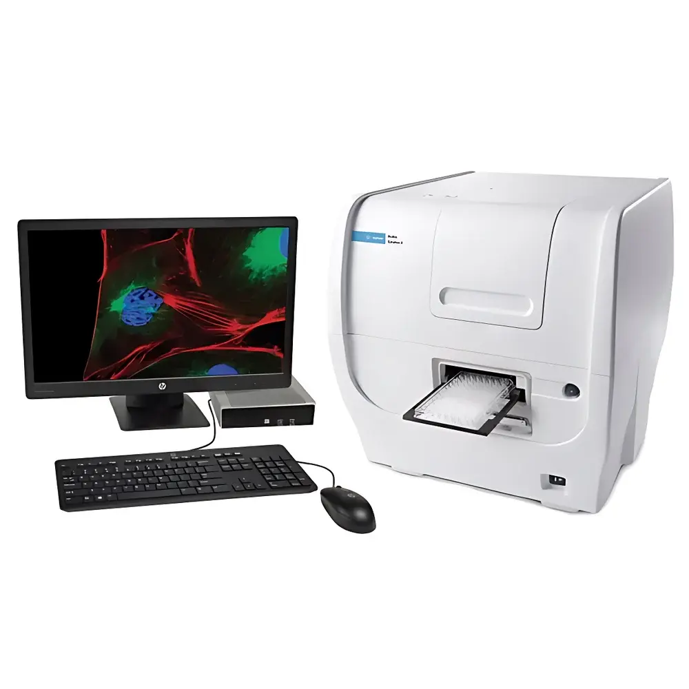
The Agilent BioTek Cytation 5 Cell Imaging Multi-Mode Reader is an integrated, modular platform engineered for quantitative high-content cellular analysis within standardized microplate formats. It combines a fully automated inverted microscope—capable of wide-field fluorescence, phase contrast, brightfield, high-contrast brightfield, and color brightfield imaging—with a high-performance multi-mode plate reader featuring dual optical paths: filter-based and quad-grating monochromator detection. This hybrid architecture enables simultaneous or sequential acquisition of morphological, spatial, and biochemical data from live or fixed cells—without requiring separate instrumentation or manual sample transfer. The system operates on the principle of digital wide-field microscopy coupled with spectrophotometric quantification, supporting kinetic, endpoint, and multiplexed assays across diverse biological contexts. Its design adheres to core requirements for GLP-compliant cell-based assay development, including traceable environmental control, audit-ready software logging, and hardware-level reproducibility validation.
Key Features
- Modular imaging architecture with six-position objective turret (1.25×–60× air objectives; 4×–40× phase contrast), enabling rapid reconfiguration for applications ranging from whole-well overview to subcellular resolution.
- Dual-path detection engine: Filter-based optics for high-sensitivity, fixed-wavelength assays (e.g., GFP, RFP, DAPI); quad-grating monochromator with adjustable bandwidth (1–50 nm) for flexible excitation/emission profiling and spectral deconvolution.
- Automated Z-stack acquisition, montage stitching, time-lapse imaging, and focus optimization via laser autofocus and motorized stage—supporting unattended long-term live-cell experiments under tightly regulated CO₂ (0–20%), O₂ (0.1–21%), and temperature (ambient to 65 °C) conditions.
- Integrated environmental control with condensation suppression and Peltier-based cooling—critical for maintaining physiological relevance during extended imaging sessions.
- High-fidelity 16-bit color CMOS camera with large field-of-view sensor, optimized for low-noise acquisition across all imaging modalities without pixel binning compromises.
- Hardware-level compatibility with Agilent’s BioSpa 8 automated incubator and BioStack plate handlers, enabling end-to-end workflow automation from plating to analysis.
Sample Compatibility & Compliance
The Cytation 5 supports standard and non-standard sample carriers—including 6–1536-well microplates, glass slides, chambered coverslips, T25 flasks, Petri dishes, hemocytometers, and Take3 microplates—ensuring seamless transition between high-throughput screening and low-throughput mechanistic studies. All imaging and detection modules comply with ISO/IEC 17025 calibration traceability frameworks when operated with documented SOPs. Environmental control subsystems meet ASTM E2917-21 specifications for thermal uniformity in incubated imaging systems. Software operation conforms to FDA 21 CFR Part 11 requirements when Gen5 is configured with electronic signatures, audit trails, and role-based access controls—making it suitable for regulated environments engaged in drug discovery, toxicology, and biomarker validation.
Software & Data Management
Gen5 v3.11+ software provides unified control over both imaging and plate reading functions, with embedded scripting (Gen5 Scripting Language), batch processing queues, and customizable analysis pipelines. Image processing includes deconvolution (Richardson-Lucy algorithm), digital phase contrast reconstruction, Z-projection (max-intensity, sum, average), and automatic montage alignment. Quantitative analysis modules support confluency measurement, nuclear/cytoplasmic segmentation, subpopulation gating (e.g., based on intensity thresholds or morphology), translocation scoring (e.g., NF-κB nuclear entry), and phenotypic profiling using machine-learning-assisted classifiers. All raw image data (TIFF, OME-TIFF), metadata (EXIF + custom tags), and analysis logs are stored in a relational database structure with version-controlled export options (CSV, PDF, XML). Data integrity is preserved through SHA-256 checksum generation and timestamped audit trails compliant with ALCOA+ principles.
Applications
- High-content phenotypic screening: Simultaneous acquisition of viability (ATP luminescence), proliferation (EdU incorporation), and morphology (nuclear size, actin organization) in single-well multiplexed assays.
- Live-cell dynamics: Real-time tracking of mitochondrial membrane potential (TMRM), calcium flux (Fluo-4), or apoptosis (Annexin V-FITC/PI) under controlled gas and thermal conditions.
- Cell migration and invasion: Automated quantification of wound healing closure rates or Transwell matrix penetration using time-lapse phase contrast and fluorescent labeling.
- Immunofluorescence and histopathology: High-resolution imaging of tissue sections or 3D spheroids with multi-channel co-localization analysis and spatial statistics (e.g., distance mapping between immune markers).
- Hit-picking and triage: Primary plate reader identification of hits (e.g., luciferase reporter activation), followed by targeted high-magnification imaging only of positive wells—reducing storage overhead and analysis time by >70% compared to full-plate imaging.
FAQ
Does the Cytation 5 support time-resolved fluorescence (TRF) and TR-FRET assays?
Yes—the system includes dedicated TRF and TR-FRET detection modes with adjustable delay times and integration windows, compatible with europium, terbium, and samarium chelates.
Can Gen5 software perform batch analysis of previously acquired images without re-acquisition?
Yes—Gen5 supports offline analysis mode, allowing users to apply updated algorithms or parameters to archived TIFF/OME-TIFF datasets while preserving original acquisition metadata.
Is CO₂ calibration required before each experiment?
No—factory-calibrated NDIR CO₂ sensors provide stable readings over 12 months; optional user-initiated zero-point verification is supported via ambient air exposure protocol.
What is the maximum supported Z-stack depth and step size?
Up to 100 focal planes per well with programmable step sizes from 0.1 µm to 100 µm, constrained by objective working distance and refractive index matching.
Does the system offer validation documentation for IQ/OQ/PQ protocols?
Agilent provides comprehensive validation templates (including test scripts, acceptance criteria, and blank execution records) aligned with ASTM E2500-13 and USP guidelines for analytical instrument qualification.


