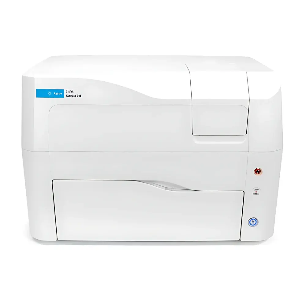
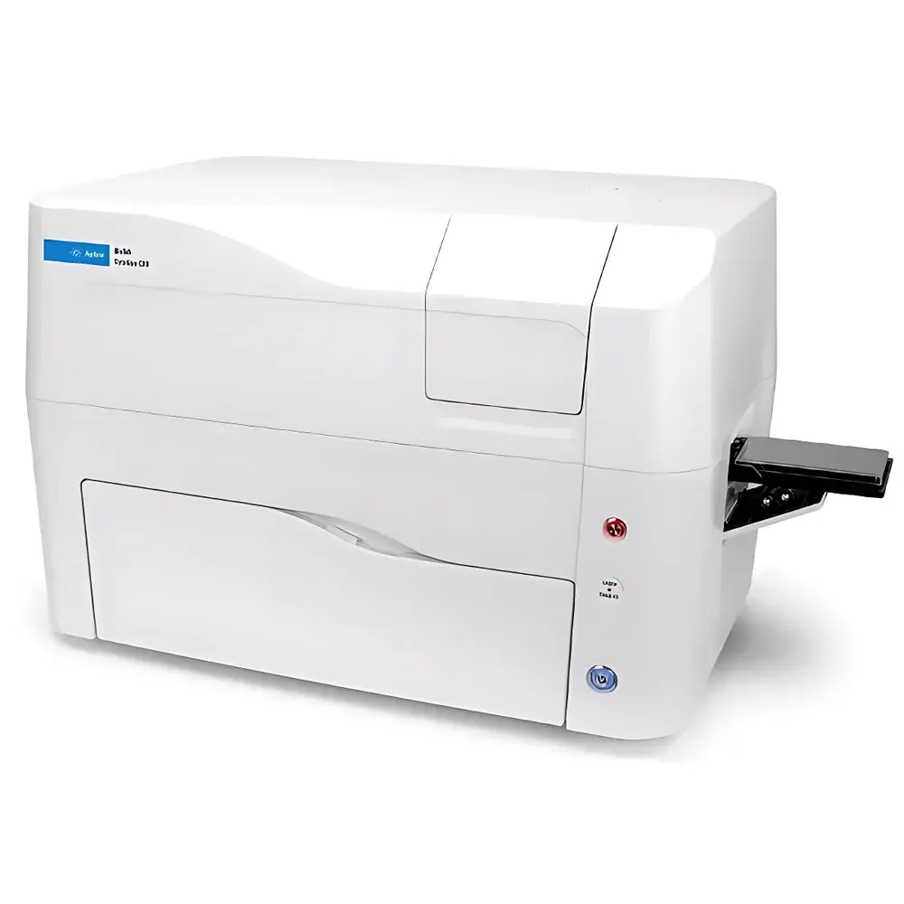
Agilent BioTek Cytation C10 Live-Cell Confocal Microplate Imaging System
| Brand | Agilent Technologies |
|---|---|
| Origin | USA |
| Manufacturer Type | Original Equipment Manufacturer (OEM) |
| Product Category | Imported Instrument |
| Model | Cytation C10 |
| Temperature Control Range | Up to 45 °C |
| Environmental Control | Dual-gas (CO₂/O₂) regulation with condensation prevention |
| Imaging Modalities | Confocal fluorescence, widefield fluorescence, brightfield, phase contrast, high-contrast brightfield, color brightfield |
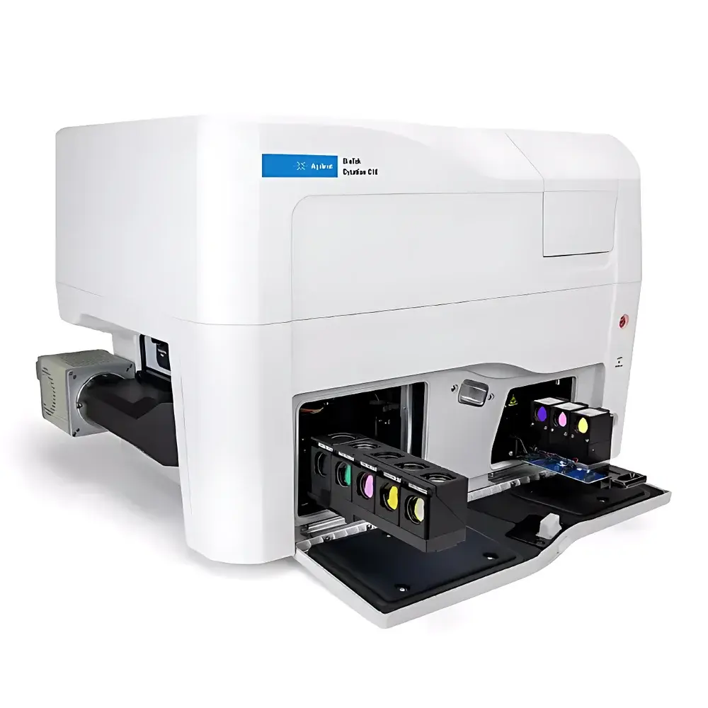
| Camera | Hamamatsu ORCA-Fusion BT sCMOS (16-bit), Sony IMX series CMOS (16-bit) |
| Objective Compatibility | Olympus air objectives (1.25×–60×), phase contrast (4×–40×), water immersion (20×–60×) |
| Plate Support | 6–1536-well microplates, slides, Petri dishes, T25 flasks, chambered coverslips, hemocytometers, Take3/Take3 Trio microvolume plates |
| Z-stack capability | Motorized focus with DSD (Deep Slice Disk) 60 µm pinhole option |
| Excitation Source | Tunable quad-grating monochromator (UV–Vis), laser-based confocal illumination |
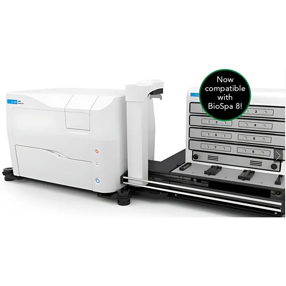
| Automation Integration | BioSpa 8 incubator, BioStack stacker |
| Software Platform | Gen5 Image+ data acquisition and analysis suite |
Overview
The Agilent BioTek Cytation C10 Live-Cell Confocal Microplate Imaging System is an integrated benchtop platform engineered for quantitative, longitudinal cellular analysis in physiologically relevant conditions. It combines dual optical pathways—spinning-disk confocal microscopy and high-sensitivity widefield imaging—within a single microplate reader architecture. The system operates on the principle of optical sectioning: the spinning-disk confocal module employs a rotating Nipkow disk with precisely aligned microlens-enhanced pinholes to reject out-of-focus light, enabling high-resolution z-stack acquisition with minimal phototoxicity. Simultaneously, the widefield path leverages high-quantum-efficiency sCMOS detection and tunable monochromator-based excitation to support rapid, multi-parametric screening across absorption, fluorescence intensity, and luminescence modes. This hybrid design bridges the gap between high-content imaging (HCI) and functional plate-based assays, allowing users to perform kinetic phenotypic profiling without compromising spatial fidelity or temporal resolution.
Key Features
- Spinning-disk confocal imaging with optional Deep Slice Disk (DSD) providing 60 µm effective optical sectioning depth—optimized for thick 3D cultures, organoids, and tissue explants.
- Water-immersion objective support (20×–60×) coupled with high-NA optics to maximize photon collection efficiency, reducing exposure time by up to 4× compared to equivalent air objectives—critical for minimizing photobleaching and photodamage during live-cell time-lapse experiments.
- Dual-camera architecture: Hamamatsu ORCA-Fusion BT sCMOS sensor (16-bit dynamic range, >95% quantum efficiency at 550 nm) for low-noise confocal acquisition; complementary Sony CMOS sensor for high-speed widefield capture.
- Four-zone independent temperature control (ambient to 45 °C) with active condensation suppression, plus programmable CO₂ (0–20%) and O₂ (1–21%) regulation—fully compliant with ISO 13485 environmental monitoring requirements for cell-based assay development.
- Tunable quad-grating monochromator (200–1000 nm) delivering <2 nm bandwidth resolution for precise spectral separation in multiplexed fluorescent assays—supporting FRET, ratiometric Ca²⁺ imaging, and spectral unmixing workflows.
- Gen5 Image+ software with automated focus algorithms (laser autofocus, contrast-based edge detection), motorized XYZ stage, and integrated montage stitching for seamless tile-based imaging across large-area samples including slides and chambered coverslips.
Sample Compatibility & Compliance
The Cytation C10 accommodates standard and non-standard formats used in translational and industrial cell biology: from 6- to 1536-well microplates, glass-bottom dishes, T25 flasks, and custom chambered substrates. Its mechanical design conforms to ANSI/SLAS standards for microplate footprint and height tolerance, ensuring interoperability with robotic liquid handlers and automated incubators such as the BioSpa 8. All environmental control subsystems are validated per ASTM E2918-21 for stability and uniformity across the sample plane. Data integrity features—including audit trail logging, user access controls, and electronic signature support—align with FDA 21 CFR Part 11 and EU Annex 11 expectations for regulated environments conducting GLP/GMP-compliant assay development.
Software & Data Management
Gen5 Image+ serves as the unified interface for instrument control, image acquisition, and quantitative analysis. It supports batch processing of multi-dimensional datasets (x, y, z, t, λ, well), with built-in modules for subcellular object segmentation (nucleus/cytoplasm/membrane), intensity normalization, colocalization (Pearson’s r, Mander’s coefficients), and kinetic curve fitting. Raw image data is stored in OME-TIFF format, preserving metadata required for MIAME/MINSEQE compliance. Export options include CSV for statistical packages (e.g., GraphPad Prism, R), ND2 compatibility for Nikon NIS-Elements, and direct integration with cloud-based analysis platforms via RESTful API. All processing steps—including thresholding, background subtraction, and deconvolution—are fully traceable and reproducible.
Applications
The Cytation C10 enables rigorous investigation across multiple life science domains: high-content toxicology (mitochondrial membrane potential, lysosomal pH, DNA damage foci); 3D tumor spheroid invasion and drug penetration kinetics; stem cell differentiation tracking using morphological and reporter-based endpoints; and immune synapse formation in co-culture models. Its ability to interleave plate-based functional readouts (e.g., ATP viability, caspase activation) with targeted confocal imaging of “hit” wells—via Gen5’s Hit-Picking workflow—reduces storage overhead by >70% versus full-plate imaging while maintaining statistical power. The system is routinely deployed in academic core facilities, biopharma discovery labs, and contract research organizations performing IND-enabling safety pharmacology studies.
FAQ
Does the Cytation C10 support long-term time-lapse imaging of primary neurons?
Yes—its four-zone thermal uniformity (<±0.3 °C), gas-regulated hypoxia capability, and low-phototoxicity water-immersion optics enable stable imaging over 72+ hours without significant drift or cellular stress.
Can I integrate third-party analysis software like Imaris or Huygens?
Yes—OME-TIFF export preserves all dimensional metadata, and Gen5 supports plugin architecture for custom algorithm injection into the acquisition pipeline.
Is the DSD 60 µm pinhole option field-upgradeable?
Yes—the spinning-disk module is modular; DSD installation requires no recalibration and is performed under Agilent Field Service supervision.
How does the system ensure focus stability during extended Z-stack acquisitions?
It combines hardware-based laser autofocus (active feedback loop) with software-driven adaptive refocusing algorithms that compensate for thermal drift and plate warping in real time.
What regulatory documentation is provided for GxP environments?
Agilent supplies IQ/OQ/PQ protocols, calibration certificates traceable to NIST standards, and a complete 21 CFR Part 11 compliance package including electronic signature validation and audit trail configuration reports.


