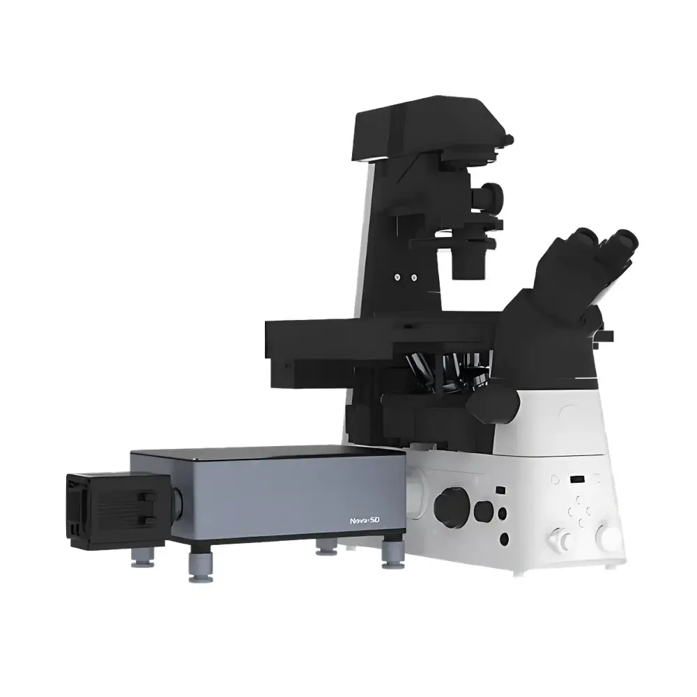
Airy Nova-SD Spinning Disk Confocal Microscopy System
| Brand | Airy |
|---|---|
| Origin | Beijing, China |
| Model | Nova-SD |
| Lateral Resolution | ~230 nm (theoretical limit: 120 nm) |
| Axial Resolution | ~600 nm (theoretical limit: 450 nm) |
| Laser Lines | 405 nm (1000 mW), 450 nm (1000 mW), 488 nm (1000 mW), 520 nm (1000 mW), 561 nm (1000 mW), 640 nm (1000 mW), 750 nm (1000 mW) |
| Detector Type | Scientific CMOS or EMCCD camera (customizable) |
| Spindle Speed | 7500 RPM |
| Frame Rate | up to 2000 fps (at reduced ROI) |
| Objective | 100× oil immersion (NA ≥ 1.4) |
| Compatible Microscope Frames | Nikon Ti2-E, Olympus IX83, Leica DMi8 |
| Illumination Sources | Same multi-wavelength laser set as excitation |
| Software & Image Workstation | Integrated acquisition and analysis suite |
| Vibration-Isolated Optical Table | Included |
| XY Stage Control | Motorized, precision piezo or stepper-driven |
Overview
The Airy Nova-SD Spinning Disk Confocal Microscopy System is a high-speed, high-sensitivity optical imaging platform engineered for live-cell and dynamic subcellular observation. Unlike point-scanning confocal systems, the Nova-SD employs a Nipkow-type spinning disk with dual microlens-enhanced architecture to simultaneously illuminate and detect multiple spatial points across the field of view. This design enables diffraction-limited optical sectioning with minimal phototoxicity and photobleaching—critical for longitudinal studies of intracellular trafficking, organelle dynamics, calcium signaling, and protein co-localization in intact tissues or cultured specimens. The system integrates seamlessly with industry-standard inverted research microscopes (Nikon Ti2-E, Olympus IX83, Leica DMi8), leveraging their mechanical stability, modular optics paths, and motorized stage compatibility. Its theoretical lateral resolution of 120 nm and axial resolution of 450 nm—achievable under optimal conditions with high-NA objectives and appropriate refractive index matching—are consistent with Abbe diffraction limits for visible-to-NIR excitation wavelengths.
Key Features
- High-throughput optical sectioning: 7500 RPM spinning disk rotation synchronized with ultra-low-latency camera readout supports real-time volumetric imaging at up to 2000 frames per second (within region-of-interest mode)
- Multi-wavelength excitation flexibility: Seven standard laser lines (405–750 nm), each delivering 1000 mW output power, enable simultaneous multi-color fluorescence acquisition with minimal crosstalk
- Modular detector integration: Supports scientific-grade sCMOS and EMCCD cameras; quantum efficiency, pixel size, and read noise are configurable per application requirement
- Precision motorized XY stage with closed-loop feedback: Enables automated tile scanning, time-lapse mosaic acquisition, and reproducible coordinate-based repositioning across experiments
- Dual-microlens disk configuration: Enhances light throughput by >3× compared to conventional single-microlens designs, improving signal-to-noise ratio without compromising resolution
- Integrated vibration isolation: Includes a passive pneumatic optical table optimized for 1 Hz, essential for long-duration Z-stack acquisition
Sample Compatibility & Compliance
The Nova-SD accommodates a broad range of biological specimens—including adherent and suspension mammalian cells, primary neurons, zebrafish embryos, Drosophila tissues, and 3D organoids—when mounted on standard #1.5 coverslips or glass-bottom dishes. It complies with ISO 10993-5 (biological evaluation of medical devices) for non-invasive imaging workflows and supports GLP-aligned experimental documentation through timestamped metadata embedding. While not FDA-cleared as an IVD device, its hardware and software architecture conform to foundational principles of 21 CFR Part 11 for audit trail generation, user access control, and electronic signature support—facilitating integration into regulated preclinical research environments.
Software & Data Management
The proprietary acquisition and analysis suite provides synchronized control of lasers, disk rotation, camera exposure, Z-focus, and stage positioning via a unified GUI. All acquired images are saved in OME-TIFF format with embedded metadata (objective NA, laser power, exposure time, pinhole diameter, Z-step interval). Batch processing pipelines support deconvolution (Richardson-Lucy algorithm), spectral unmixing (linear unmixing with reference spectra), and intensity normalization across time series. Raw data export supports HDF5 and N5 container formats for compatibility with Python-based analysis frameworks (e.g., Napari, CellProfiler, BigDataViewer). Audit logs record operator ID, session start/end timestamps, parameter modifications, and file export events—enabling traceability in multi-user core facilities.
Applications
- Live-cell calcium imaging using GCaMP or Fluo-4 dyes at sub-second temporal resolution
- Endosomal and lysosomal trafficking assays with dual-channel labeling (e.g., Rab5/Rab7)
- Microtubule and actin cytoskeleton dynamics during mitosis or migration
- Co-localization quantification in fixed samples using Pearson’s correlation and Manders’ coefficients
- 3D reconstruction of cleared tissue sections (CLARITY, iDISCO) with isotropic voxel sampling
- High-content screening of siRNA or CRISPR perturbations in 96-/384-well plates
FAQ
What microscope frames are natively supported?
The Nova-SD is mechanically and optically calibrated for Nikon Ti2-E, Olympus IX83, and Leica DMi8 inverted platforms. Custom adapter kits are available for other models upon request.
Can the system perform resonant scanning or line scanning?
No—the Nova-SD is exclusively a spinning disk confocal platform. Resonant or galvo-based scanning modes are not implemented.
Is the software compatible with third-party analysis tools like Imaris or Arivis Vision4D?
Yes. Exported OME-TIFF files retain full dimensional metadata and can be directly imported into commercial visualization and quantification platforms.
What is the maximum usable field of view at 100× magnification?
With a 22-mm camera sensor and 100×/1.4 NA objective, the FOV is approximately 120 µm × 120 µm (full frame), scalable via binning or ROI selection.
Does the system include temperature and CO₂ control integration?
Environmental chamber interfaces (for stage-top incubators or enclosure-based systems) are available as optional accessories and support TTL/RS232 communication protocols.


