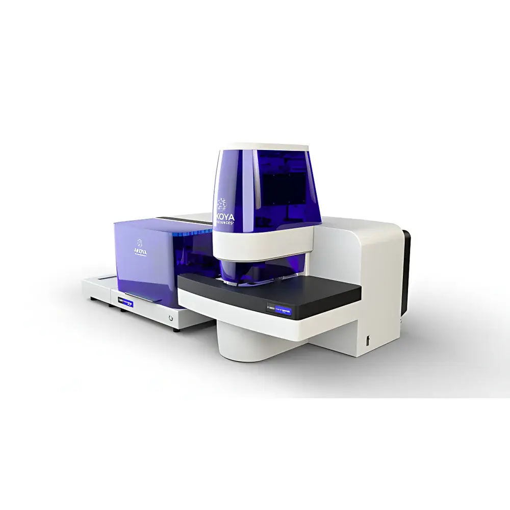
Akoya Biosciences PhenoCycler-Fusion Single-Cell In Situ Spatial Omics Analysis Platform
| Brand | Akoya |
|---|---|
| Origin | USA |
| Model | PhenoCycler-Fusion |
| Detection Modality | Cyclic Immunofluorescence (CyCIF) + In Situ Hybridization (ISH)-compatible |
| Pixel Resolution | 0.25 µm/pixel |
| Throughput | 5–100 tissue sections per week |
| Multiplex Capacity | >100 protein biomarkers per run |
| Imaging Speed | ~1 million cells imaged in ≤10 minutes |
| Compatible Analytes | Proteins and RNA in formalin-fixed paraffin-embedded (FFPE) and frozen tissue sections |
Overview
The Akoya Biosciences PhenoCycler-Fusion is an integrated, high-throughput spatial omics platform engineered for single-cell-resolution, in situ molecular profiling of intact tissue sections. It leverages cyclic immunofluorescence (CyCIF) combined with iterative antibody stripping and re-probing—augmented by optional in situ hybridization (ISH) modules—to enable simultaneous, quantitative detection of over 100 protein targets and/or RNA species within native tissue architecture. Unlike conventional IHC or sequential IF platforms, PhenoCycler-Fusion employs a closed-loop, fully automated fluidic and imaging workflow that eliminates manual intervention between cycles, ensuring consistent antigen retrieval, antibody binding kinetics, and signal quantification across all rounds. Its optical design supports sub-micron pixel resolution (0.25 µm/pixel), enabling precise cellular segmentation and spatial neighbor analysis at true single-cell granularity. The system is optimized for both FFPE and fresh-frozen human and murine tissue sections, supporting reproducible, GLP-aligned spatial phenotyping workflows required for translational research and biomarker discovery.
Key Features
- Ultra-High Multiplexing: Detects ≥100 protein biomarkers per tissue section using validated, spectrally resolved antibody panels—compatible with Akoya’s CODEX®-certified reagents and third-party conjugated antibodies meeting CyCIF specifications.
- Single-Cell Spatial Fidelity: Achieves 0.25 µm/pixel resolution via precision motorized stage control and high-numerical-aperture objective optics, enabling accurate nuclear/cytoplasmic compartmentalization and subcellular feature extraction.
- Accelerated Workflow: Acquires spatially registered, multi-parametric data from ~1 million cells in under 10 minutes per cycle—reducing total assay time by up to 70% compared to legacy cyclic platforms.
- End-to-End Automation: Integrates on-instrument antibody labeling, washing, stripping, imaging, and autofocus calibration—minimizing operator variability and supporting unattended overnight operation.
- Multi-Omic Flexibility: Supports parallel or sequential detection of proteins and RNA targets on the same section using orthogonal probe chemistries (e.g., tyramide signal amplification for proteins; branched DNA or hybridization chain reaction for RNA).
- Robust Architecture: Built with ISO 13485-aligned manufacturing standards; includes hardware-level environmental monitoring (temperature, humidity, vibration damping) to maintain imaging stability across multi-day acquisitions.
Sample Compatibility & Compliance
PhenoCycler-Fusion accepts standard 1–4 µm thick tissue sections mounted on charged glass slides (e.g., Superfrost Plus), compatible with routine histopathology workflows. It supports FFPE blocks processed using standard clinical protocols—including automated deparaffinization and epitope retrieval—and frozen sections prepared via cryostat without antigen masking artifacts. All reagent kits are supplied with Certificate of Analysis (CoA) and lot-specific validation data aligned with CLIA- and CAP-recommended controls. The platform complies with FDA 21 CFR Part 11 requirements for electronic records and signatures when used with Akoya’s validated software stack, and supports audit trails, user access controls, and versioned protocol archiving for GCP/GLP/GMP-regulated studies.
Software & Data Management
Acquisition and analysis are managed through Phenoptics™ Software Suite v6.0+, which provides instrument control, real-time QC metrics (signal-to-noise ratio, cycle-to-cycle registration error, autofluorescence baseline), and integrated AI-assisted cell segmentation (using U-Net-based deep learning models trained on >50,000 annotated tissue regions). Raw TIFF stacks are stored in OME-TIFF format compliant with the Open Microscopy Environment (OME) standard. Export options include HDF5 for downstream spatial transcriptomics integration (e.g., with Seurat, Squidpy, or SpatialData), CSV for statistical modeling, and MCD/IMC-compatible .txt for cross-platform comparison. Data integrity is enforced via SHA-256 checksum verification at ingestion and automatic backup to network-attached storage (NAS) or cloud repositories (AWS S3, Azure Blob) with configurable retention policies.
Applications
- De novo construction of spatially resolved human tissue atlases (e.g., tumor microenvironment mapping, lymphoid organ zoning, neuroanatomical layering)
- Discovery of rare or transitional cell states in disease progression (e.g., pre-metastatic niches, therapy-resistant clones, senescence-associated secretory phenotypes)
- Validation of spatial biomarkers for companion diagnostics—aligned with CAP/ISO 15189 preanalytical and analytical validation frameworks
- Integration with bulk or single-cell RNA-seq datasets to resolve expression-context discordance (e.g., ligand–receptor co-expression in stromal–immune crosstalk)
- Longitudinal spatial profiling of treatment response in PDX or humanized mouse models under controlled vivarium conditions
FAQ
What tissue preparation methods are validated for use with PhenoCycler-Fusion?
Standard FFPE processing (including automated stainer-derived sections) and OCT-embedded frozen sections are fully supported. Antigen retrieval must follow Akoya-specified pH- and temperature-controlled protocols.
Can PhenoCycler-Fusion be used for clinical trial sample analysis?
Yes—when operated under validated SOPs and paired with Akoya’s IQ-Verify™ quality assurance module, the system meets analytical validity requirements for exploratory biomarker analysis in Phase I–III trials.
Is raw image data export compliant with FAIR principles?
Yes—OME-TIFF metadata includes MIAME/MINSEQE-compliant descriptors, instrument configuration logs, and reagent lot traceability, enabling Findable, Accessible, Interoperable, and Reusable data stewardship.
How does the platform handle autofluorescence in highly pigmented or aged tissues?
Built-in spectral unmixing algorithms (linear least-squares fitting against reference spectra) and adaptive background subtraction are applied per cycle, with optional dark-field illumination mode for melanin-rich samples.
What level of IT infrastructure is required for local deployment?
Minimum: 10 GbE network, RAID-6 NAS with ≥100 TB usable capacity, and Windows Server 2022 with 128 GB RAM and dual NVIDIA A100 GPUs for accelerated segmentation.
Related Products


