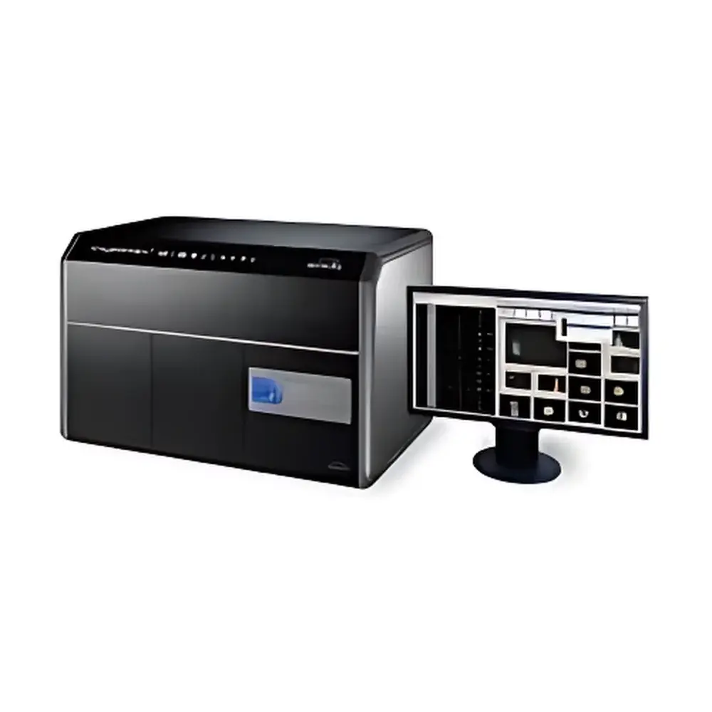
Amnis ImageStreamX Mark II Imaging Flow Cytometer
| Brand | Amnis |
|---|---|
| Origin | USA |
| Manufacturer | Luminex Corporation (acquired Amnis) |
| Product Type | Imaging Flow Cytometer |
| Model | ImageStreamX Mark II |
| Laser Configuration | Up to 7 lasers (standard: 488 nm, 642 nm, 785 nm |
| optional | 375 nm, 405 nm, 561 nm, 592 nm) |
| Detection Channels | Standard: 6 channels (1 brightfield, 1 darkfield, up to 4 fluorescence) |
| Objective Lenses | Standard 40× |
| Fluorescence Sensitivity | <5 MESF (Molecules of Equivalent Soluble Fluorochrome) |
| Imaging Rate | >1,000 cells/sec |
| Images per Cell | Up to 12 simultaneous high-resolution digital images (including BF, DF, and multiple fluorescence channels) |
| Pixel Resolution | 656 × 494 pixels per image |
| Field of View | ~100 µm × 75 µm per image |
| Sample Volume Range | 20–200 µL per acquisition |
Overview
The Amnis ImageStreamX Mark II is a second-generation imaging flow cytometer engineered to bridge the analytical rigor of conventional flow cytometry with the morphological and spatial resolution of fluorescence microscopy. Unlike traditional flow cytometers that report only scalar fluorescence intensities and light-scatter parameters, the ImageStreamX Mark II captures high-fidelity, multi-channel digital images of every cell in suspension—enabling quantitative analysis of morphology, subcellular localization, co-localization, nuclear translocation, phagocytosis, cell–cell interactions, and other functional phenotypes at single-cell resolution. Its core measurement principle relies on hydrodynamic focusing combined with time-delay integration (TDI) CCD imaging, synchronized with laser excitation and precise fluidic control. This architecture delivers statistically robust, high-throughput image-based cytometry data compatible with standard flow cytometry workflows while extending analytical depth into spatial and contextual dimensions previously inaccessible in population-level assays.
Key Features
- Simultaneous acquisition of up to 12 digital images per cell—including brightfield (BF), darkfield (DF), and up to 10 fluorescence channels—captured at >1,000 cells per second.
- Modular laser architecture supporting up to seven solid-state lasers (375, 405, 488, 561, 592, 642, 785 nm), enabling broad spectral coverage for multicolor panel design and advanced applications such as FRET, spectral unmixing, and deep-tissue-compatible NIR imaging.
- High-sensitivity detection system with <5 MESF sensitivity across all fluorescence channels, validated using standardized calibration beads traceable to NIST reference materials.
- Interchangeable objective turret accommodating 20×, 40×, and 60× air objectives—each optimized for specific balance between field-of-view, resolution, and signal-to-noise ratio—allowing flexible experimental design from large-particle screening to subcellular detail analysis.
- Integrated fluidics with precision syringe pump and pressure-controlled sample introduction, ensuring stable flow rates and minimal shear stress—critical for fragile primary cells, organoids, or rare circulating tumor cells (CTCs).
- Robust mechanical and thermal design compliant with ISO 13485–certified manufacturing standards; CE-marked and FDA 510(k)-cleared for clinical research use in IVD-limited settings.
Sample Compatibility & Compliance
The ImageStreamX Mark II accepts a wide range of biological samples without fixation or mounting requirements: unfixed or fixed suspension cells (e.g., PBMCs, leukemic blasts), adherent cells detached via enzymatic or non-enzymatic methods, yeast, bacteria, extracellular vesicles, and even small multicellular aggregates (<50 µm). It supports standard flow cytometry tubes (12 × 75 mm), microcentrifuge tubes (0.5–1.5 mL), and custom sample reservoirs. Data acquisition and analysis comply with GLP and GMP-aligned documentation practices. IDEAS® software includes full audit trail functionality, electronic signature support, and export formats compatible with 21 CFR Part 11 requirements for regulated environments. Instrument performance verification follows ASTM E2877-22 (Standard Guide for Validation of Imaging Flow Cytometers) and ISO/IEC 17025–aligned protocols.
Software & Data Management
Data acquisition and analysis are performed using IDEAS® software—a purpose-built platform integrating image processing, machine learning–assisted feature extraction, and flow-like gating logic. IDEAS provides over 100 pre-defined, biologically relevant features—including object area, intensity, texture, shape descriptors (e.g., circularity, aspect ratio), proximity metrics (e.g., nuclear-cytoplasmic ratio, internalized probe distance), and co-localization coefficients (Pearson’s r, Mander’s overlap). Batch processing, template-based analysis, and customizable scripting (via Python API) enable reproducible, high-throughput quantification across hundreds of samples. Raw image data are stored in standardized .ims format (based on TIFF), preserving pixel-level metadata including exposure time, laser power, gain, and calibration references—ensuring full traceability and reanalysis capability years after acquisition.
Applications
- Cellular Functional Phenotyping: Quantification of NF-κB nuclear translocation, p53 activation kinetics, mitochondrial membrane potential redistribution, and caspase cleavage patterns under pharmacologic or genetic perturbation.
- Rare Event Detection: Identification and morphometric characterization of circulating tumor cells (CTCs), minimal residual disease (MRD) cells, or antigen-specific T-cell clones in heterogeneous backgrounds.
- Host–Pathogen Interactions: High-content analysis of bacterial internalization, viral entry mechanisms, and intracellular replication compartments using multi-channel co-localization and temporal tracking.
- Immunology & Inflammation: Discrimination of macrophage polarization states (M1/M2), neutrophil extracellular trap (NET) formation, and B-cell receptor clustering dynamics.
- Stem Cell & Regenerative Medicine: Monitoring differentiation trajectories via morphological entropy, organelle reorganization, and lineage marker spatial segregation in heterogeneous progenitor populations.
FAQ
What distinguishes the ImageStreamX Mark II from conventional flow cytometers?
It combines quantitative fluorescence intensity measurements with high-resolution digital imaging of every cell in real time—enabling morphometric, spatial, and contextual analysis not possible with scatter-only or intensity-only platforms.
Is IDEAS® software included with the instrument purchase?
Yes—IDEAS® v6.x or later is supplied with full licensing, including all core modules, batch analysis tools, and regulatory-compliant audit trail functionality.
Can the system be integrated into automated liquid handling workflows?
Yes—via RS-232 and Ethernet interfaces, the ImageStreamX Mark II supports bidirectional communication with third-party automation platforms (e.g., Hamilton STAR, Tecan Freedom EVO) for walk-away sample loading and acquisition scheduling.
What maintenance is required to ensure long-term optical alignment stability?
Annual preventive maintenance by Luminex-certified field service engineers is recommended; the system includes built-in alignment diagnostics and auto-calibration routines for daily QC verification.
Are there published validation studies demonstrating reproducibility across laboratories?
Yes—over 150 peer-reviewed publications (including in Nature Methods, Journal of Immunology, and Cytometry A) report inter-laboratory concordance of ImageStream-derived metrics using standardized SOPs and reference controls.


