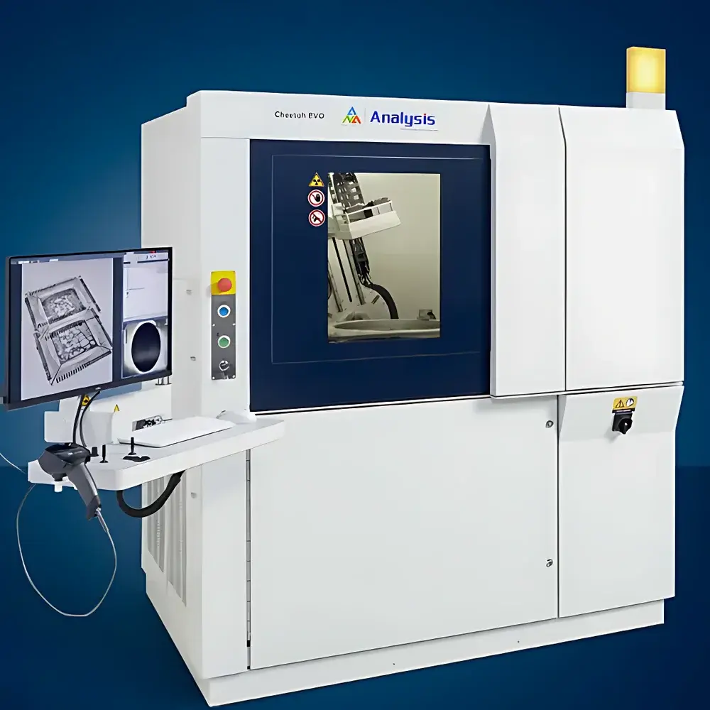
Analysis Cheetah EVO Microfocus X-ray Imaging & Industrial Micro-CT System
| Brand | Analysis |
|---|---|
| Origin | Germany |
| Model | Cheetah EVO |
| Detector Type | Flat-panel Detector |
| Scan Mode | Translation-Rotation (TR) |
| Spatial Resolution | Up to 2.5 lp/mm |
| X-ray Energy | 160 kV |
| Penetration Capability | Up to 350 mm steel-equivalent thickness |
| Precision | Nanoscale |
| Optional Features | Water-cooled X-ray tube (FXT 160.51), High-load capacity (< 20 kg), Low-dose imaging mode, Dose-reduction kit for sensitive components |
Overview
The Analysis Cheetah EVO is a high-performance microfocus X-ray imaging and industrial micro-computed tomography (micro-CT) system engineered for non-destructive evaluation (NDE) in precision manufacturing and advanced R&D environments. Operating on the principle of cone-beam microfocus radiography and iterative reconstruction-based CT, the system delivers quantitative 2D radiographic and volumetric 3D datasets with nanoscale geometric fidelity. Its 160 kV sealed-tube X-ray source—optionally available with water-cooled FXT 160.51 configuration—maintains stable focal spot positioning (< 1 µm drift over >8-hour continuous operation), eliminating thermal-induced image degradation common in air-cooled systems. This stability underpins measurement repeatability critical for ISO/IEC 17025-accredited laboratories and GMP-regulated production validation. The system complies with IEC 61331-1 (radiation protection) and meets electromagnetic compatibility requirements per EN 61326-1, ensuring safe integration into controlled cleanroom and factory-floor settings.
Key Features
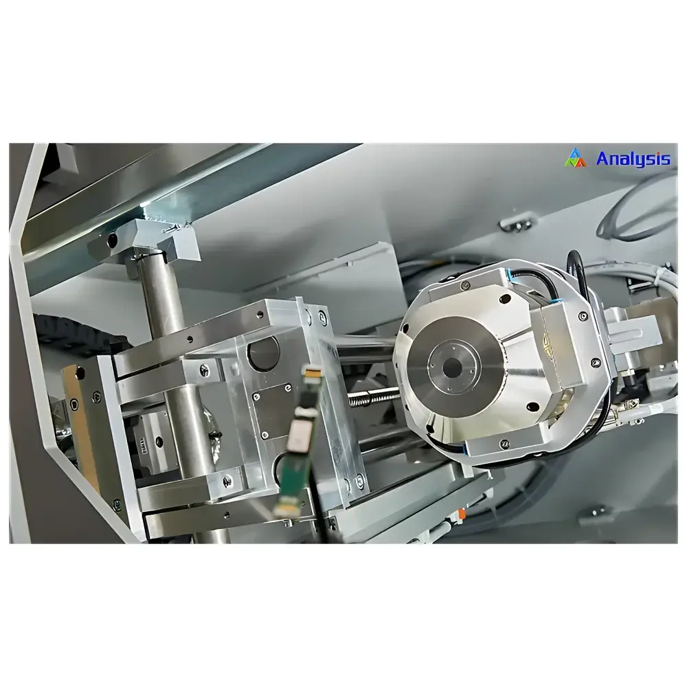
- Microfocus X-ray source with stable sub-micron focal spot, enabled by optional water-cooling architecture for extended-duty-cycle scanning without focal drift
- Large-area flat-panel detector enabling up to 50% larger field-of-view (FOV) vs. prior-generation systems—reducing multi-position stitching and accelerating throughput
- Translation-Rotation (TR) scanning geometry optimized for high-fidelity CT reconstruction, supporting both step-and-shoot and continuous acquisition modes
- Integrated low-dose imaging suite including dose-reduction hardware kit and software-controlled exposure modulation—validated for inspection of radiation-sensitive semiconductor packages and polymer-based medical devices
- FGUI (Flexible Graphical User Interface) platform with pre-configured, audit-trail-enabled workflows compliant with FDA 21 CFR Part 11 requirements for electronic records and signatures
- ProLoop communication interface enabling bidirectional data exchange with AOI/AXI platforms and MES/SCADA systems—supporting Industry 4.0 traceability and real-time SPC feedback loops
Sample Compatibility & Compliance
The Cheetah EVO accommodates samples ranging from miniature IC packages (<5 mm) to mid-size automotive castings (up to 300 mm diameter × 250 mm height), with optional high-load mechanical stage supporting static loads ≤20 kg. It supports defect detection thresholds down to <1 µm voids in solder joints, <50 µm delaminations in multilayer PCBs, and sub-100 µm porosity clusters in additively manufactured metal parts. All system firmware and FF CT reconstruction algorithms are validated against ASTM E1441 (Standard Guide for Computed Tomography), ISO 15739 (Imaging system noise metrics), and USP <1059> (Radiographic testing of pharmaceutical packaging). Full GLP/GMP documentation packages—including IQ/OQ/PQ protocols, calibration certificates traceable to PTB (Physikalisch-Technische Bundesanstalt), and change control logs—are available upon request.
Software & Data Management
The system ships with two tightly integrated software suites: FGUI for acquisition control and FF CT for reconstruction and analysis. FF CT implements GPU-accelerated Feldkamp-Davis-Kress (FDK) and iterative SART algorithms, delivering isotropic voxel resolutions down to 0.5 µm³ with contrast-to-noise ratio (CNR) >15 dB in low-contrast regions. Its proprietary “micro3Dslice” engine enables slice-by-slice quantification of internal structures without manual segmentation. The VoidInspect module performs automated void volume, sphericity, and spatial distribution analysis on BGA/CSP solder joints—outputting ASME Y14.5-compliant GD&T reports. All visualization employs cinematic rendering via pre-defined transfer functions (TFs), generating publication-ready 3D movies with depth-cued transparency and surface shading. Raw projection data and reconstructed volumes are stored in DICOM 3.0-compliant format with embedded metadata (acquisition parameters, calibration IDs, operator credentials), ensuring interoperability with PACS and LIMS infrastructure.
Applications
- SMT & semiconductor manufacturing: Non-destructive verification of flip-chip underfill integrity, wire bond lift-off, TSV (through-silicon via) fill uniformity, and Cu pillar void content—per IPC-A-610 and J-STD-001 standards
- Aerospace & automotive: Porosity mapping in high-pressure die-cast aluminum housings, fiber orientation analysis in CFRP composites, and cooling channel verification in turbine blades
- Medical device R&D: Structural validation of laser-welded implant housings, electrode coating thickness distribution in Li-ion battery cells, and microchannel geometry in microfluidic diagnostic cartridges
- Academic & national lab research: In situ mechanical testing (tensile/compression) inside the X-ray chamber using modular load frames, coupled with time-resolved 4D-CT reconstruction
FAQ
What is the minimum detectable defect size in solder joints?
The system achieves reliable detection of voids ≥1 µm in diameter within SnAgCu solder under standard acquisition conditions (160 kV, 100 µA, 1200 ms exposure); performance scales with magnification and signal averaging.
Does the Cheetah EVO support automated pass/fail classification per IPC-A-610?
Yes—via FGUI-integrated rule-based defect classification engines trained on certified reference standards; configurable acceptance criteria align with Class 2 and Class 3 requirements.
Can raw projection data be exported for third-party reconstruction?
Yes—uncompressed TIFF stacks and HDF5-formatted sinograms are exportable with full metadata, supporting custom algorithm development in MATLAB, Python (scikit-image, tomopy), or commercial platforms like Avizo.
Is remote operation and monitoring supported?
The system includes secure TLS-encrypted VNC access, RESTful API endpoints for job queue management, and SNMP v3 integration for network-wide health monitoring—fully compatible with IT-managed infrastructure.
How is geometric calibration maintained over time?
A motorized calibration phantom with traceable tungsten markers is used during scheduled OQ; automated centroid detection and polynomial correction models are applied in real time during reconstruction to compensate for mechanical drift.


