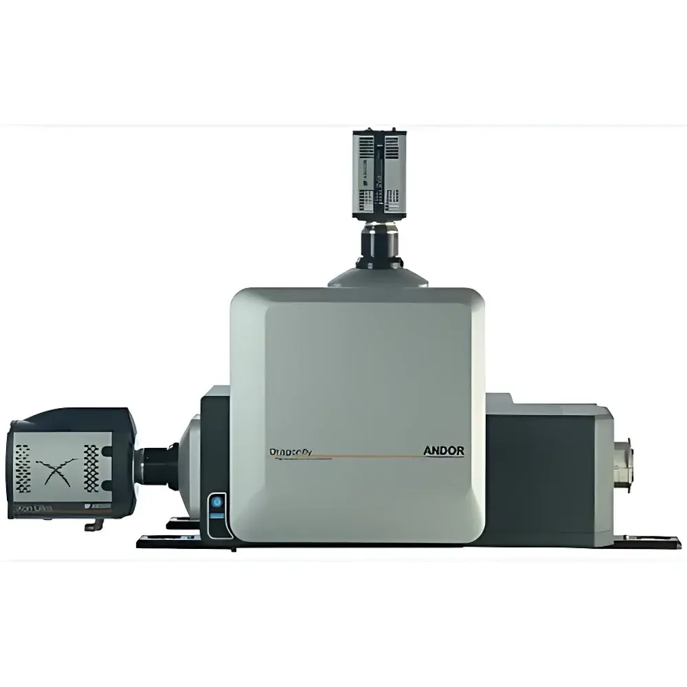
Andor Dragonfly Confocal Imaging Platform by Oxford Instruments
| Brand | Oxford Instruments |
|---|---|
| Origin | United Kingdom |
| Model | Andor Dragonfly |
| Software | Imaris (3D/4D image analysis), Fusion (acquisition & processing) |
| Imaging Modes | Laser Scanning Confocal, Widefield + GPU-Accelerated Deconvolution (ClearView-GPU™), TIRF (chromatically corrected), HILO, SRRF-Stream®, dSTORM, Iterative Deconvolution |
| Max Frame Rate | 400 fps (Dragonfly 200 series) |
| Detector Compatibility | EMCCD and sCMOS cameras |
| Core Technology | Microlens-based parallel confocal scanning head with Borealis™ uniform illumination |
Overview
The Andor Dragonfly Confocal Imaging Platform is a high-performance, modular optical imaging system engineered by Oxford Instruments for advanced fluorescence microscopy across life sciences research. Unlike conventional point-scanning confocal microscopes, the Dragonfly employs a patented microlens-based parallel confocal scanning architecture—integrated directly with high-sensitivity EMCCD or sCMOS detectors—to deliver up to 20× faster volumetric acquisition without compromising signal fidelity. Its core design eliminates mechanical scanning delays, enabling true real-time 3D visualization of dynamic biological processes—from single-molecule localization to whole-embryo developmental imaging. The platform supports multiple complementary modalities within a single optical path: laser scanning confocal, widefield fluorescence with CUDA-GPU-accelerated ClearView-GPU™ deconvolution, chromatically corrected total internal reflection fluorescence (TIRF), HILO (highly inclined laminated optical sheet), and super-resolution techniques including SRRF-Stream® and dSTORM. This multi-modal flexibility allows researchers to select the optimal imaging strategy per sample thickness, labeling density, photostability, and temporal resolution requirement—without hardware reconfiguration.
Key Features
- Parallel Confocal Architecture: Patented microlens array enables simultaneous multi-point illumination and detection, achieving frame rates up to 400 fps in confocal mode—significantly exceeding conventional galvo- or resonant-scanning systems.
- Borealis™ Uniform Illumination: Engineered for large-field homogeneity across objectives up to 25 mm FOV, ensuring quantitative intensity consistency in tiled acquisitions and thick-tissue imaging.
- GPU-Accelerated Processing: ClearView-GPU™ deconvolution operates at 10–20× the speed of CPU-based algorithms, enabling near-real-time restoration of widefield data with sub-diffraction contrast enhancement.
- Multi-Modal Flexibility: Seamless switching between confocal, widefield, TIRF, HILO, and super-resolution modes via software control; all share the same optical train and detector interface.
- Dual-Camera Acquisition Support: Enables simultaneous spectral or temporal channel separation—for example, ratiometric Ca²⁺ imaging or FRET with independent gain and exposure optimization per channel.
- Automated Focus Maintenance: Integrated hardware autofocus maintains Z-position stability during long-term time-lapse or multi-position experiments, critical for reproducible 4D datasets.
Sample Compatibility & Compliance
The Dragonfly platform accommodates a broad range of specimen types—from live single cells and organoids to cleared whole-mount tissues and zebrafish embryos. Its adjustable TIRF angle control (0.1° resolution) and HILO illumination depth tuning allow precise excitation confinement for membrane-proximal or cytoplasmic targets while minimizing out-of-focus background. For thick samples (>100 µm), the combination of high quantum efficiency sCMOS/EMCCD detection, Borealis™ illumination uniformity, and iterative GPU-accelerated deconvolution ensures robust signal recovery and axial sectioning capability. The system complies with ISO 10993 biocompatibility standards for optical components in contact with biological media and meets CE marking requirements for laboratory instrumentation. Data acquisition workflows support audit-trail logging and metadata embedding in OME-TIFF format—fully compatible with GLP/GMP-aligned imaging pipelines and FDA 21 CFR Part 11–compliant environments when deployed with validated Imaris and Fusion software configurations.
Software & Data Management
Acquisition and real-time processing are managed through Fusion software—a unified interface supporting hardware synchronization, multi-modal protocol scripting, and on-the-fly GPU deconvolution. Fusion exports native OME-TIFF files containing complete metadata (objective, laser power, pinhole size, z-step, camera settings), ensuring FAIR (Findable, Accessible, Interoperable, Reusable) data principles. Imaris provides advanced post-processing: automated surface rendering, filament tracing, spot detection at sub-pixel precision, and colocalization quantification across 3D/4D volumes. Both Fusion and Imaris support batch processing pipelines, Python API integration (ImarisXT), and direct export to HDF5 or N5 formats for AI-driven segmentation training. All software modules undergo annual validation against NIST-traceable reference standards for intensity linearity, spatial calibration, and photobleaching correction accuracy.
Applications
- Live-Cell Dynamics: High-speed confocal imaging of mitochondrial fission/fusion, vesicle trafficking, or calcium wave propagation at >100 fps with minimal phototoxicity.
- Super-Resolution Structural Biology: SRRF-Stream® for live-cell nanoscale imaging (~60 nm resolution) and dSTORM for fixed-sample molecular mapping (<20 nm lateral precision).
- Neuroscience: Whole-brain clearing-compatible imaging of synaptic protein distribution using multi-color TIRF/HILO/confocal fusion in thick sections.
- Developmental Biology: Long-term 4D tracking of cell lineage in zebrafish or Drosophila embryos with adaptive focus maintenance and low-photodamage illumination.
- Quantitative Drug Screening: Multiplexed nuclear/cytoplasmic/membrane marker analysis in 384-well plates using automated stage control and batch deconvolution in Fusion.
FAQ
What distinguishes Dragonfly from traditional point-scanning confocal systems?
Dragonfly uses parallel confocal detection via a microlens array instead of sequential point scanning—enabling orders-of-magnitude faster volumetric acquisition while preserving optical sectioning and signal-to-noise ratio.
Can Dragonfly perform both confocal and super-resolution imaging on the same sample?
Yes. Through software-selectable modalities—including SRRF-Stream®, dSTORM, and iterative deconvolution—the system acquires and processes data across resolution regimes without physical hardware changes.
Is Imaris included with the system, or licensed separately?
Imaris is supplied as a perpetual license with optional annual maintenance for updates and technical support; Fusion acquisition software is bundled with all Dragonfly configurations.
Does Dragonfly support objective changers and motorized stages?
Yes. It integrates natively with major microscope manufacturers’ automation APIs (Nikon NIS-Elements, Olympus cellSens, Zeiss ZEN) for synchronized stage, focus, filter, and objective control.
What level of technical support and application assistance is provided post-installation?
Oxford Instruments offers on-site installation, operator training, and dedicated application scientist support—including experimental design consultation and custom macro development for Fusion and Imaris.


