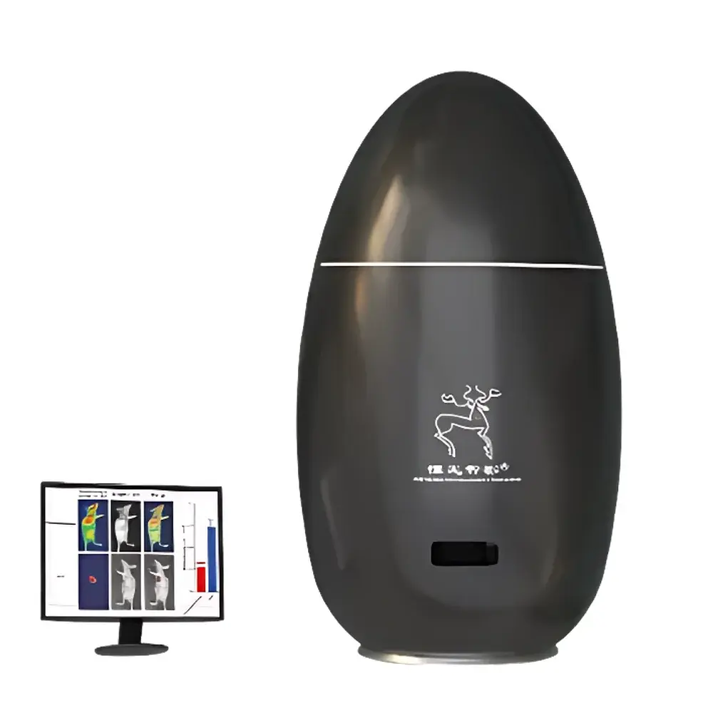
Artemis MARS Small Animal In Vivo Imaging System
| Brand | Artemis |
|---|---|
| Model | MARS |
| Origin | Shanghai, China |
| Manufacturer Type | Original Equipment Manufacturer (OEM) |
| Imaging Modalities | Fluorescence (400–900 nm), X-ray, Computed Tomography (CT) |
| Category | Optical & Multimodal Preclinical Imaging System |
| Regulatory Classification | Class II Medical Device (China NMPA), CE-marked for research use only (RUO) |
Overview
The Artemis MARS Small Animal In Vivo Imaging System is a high-performance, multimodal preclinical imaging platform engineered for quantitative, non-invasive longitudinal monitoring of biological processes in live rodents. It integrates three complementary imaging modalities—wide-spectrum fluorescence (400–900 nm), high-resolution digital radiography (X-ray), and micro-CT—within a single, compact gantry architecture. Unlike conventional optical-only systems, the MARS platform leverages co-registered multimodal acquisition to resolve anatomical context (via X-ray/CT) alongside functional or molecular contrast (via near-infrared fluorescence). Its optical path employs cooled scientific CMOS detection with quantum efficiency >80% at 700 nm, enabling high signal-to-noise ratio imaging at low photon flux—critical for longitudinal studies requiring minimal phototoxicity and dose-controlled excitation. The system operates under controlled environmental conditions (integrated temperature and gas regulation) to maintain physiological stability during extended acquisitions, supporting compliance with GLP-aligned experimental protocols.
Key Features
- Multimodal core architecture: Synchronized fluorescence, X-ray, and CT imaging in one instrument—eliminating inter-system registration errors and reducing animal handling time.
- Optical performance: 12-bit dynamic range, motorized zoom lens (0.8×–4.0×), automated focus and exposure calibration, and spectral unmixing capability for up to four fluorophores (e.g., Cy5.5, ICG, IRDye800CW, AlexaFluor750).
- X-ray subsystem: 40 kVp microfocus source, 50 µm focal spot size, 100 µm spatial resolution at detector plane; supports both projection radiography and fast cone-beam CT reconstruction (≤60 s per full 360° scan).
- CT imaging: Isotropic voxel resolution down to 50 µm; quantitative Hounsfield unit (HU)-calibrated reconstruction using NIST-traceable phantoms; supports bone, soft-tissue, and contrast-enhanced segmentation.
- Animal handling: Integrated heated stage with real-time rectal temperature feedback, isoflurane anesthesia delivery port, and adjustable positioning cradle compatible with mice (10–35 g) and rats (100–500 g).
- Robust mechanical design: Vibration-damped optical table base, EMI-shielded enclosure, and ISO Class 5 cleanroom-compatible housing for GMP-aligned facility integration.
Sample Compatibility & Compliance
The MARS system is validated for use with standard laboratory rodent models including C57BL/6, BALB/c, nude, NSG, and humanized mouse strains. It accommodates fluorescent probes compliant with FDA-approved or investigational new drug (IND)-enabling labeling (e.g., indocyanine green, bevacizumab-IRDye800CW, anti-CD31-Cy5.5). All imaging protocols adhere to ARRIVE 2.0 guidelines and support alignment with NIH Office of Laboratory Animal Welfare (OLAW) standards. Data output formats comply with DICOM 3.0 (for X-ray/CT) and MIAME-compliant TIFF/NRRD (for fluorescence), facilitating integration into institutional PACS or preclinical imaging repositories. The system meets IEC 61000-6-3 (EMC) and IEC 61000-6-2 (immunity) requirements and carries CE marking for research use only (RUO)—not intended for diagnostic or clinical applications.
Software & Data Management
Acquisition and analysis are managed via Artemis ImageSuite v4.2—a modular, audit-trail-enabled software platform compliant with 21 CFR Part 11 requirements for electronic records and signatures. Core modules include AutoAlign (rigid + deformable multimodal registration), QuantStudio (ROI-based intensity quantification with background subtraction and bleed-through correction), and TomoRecon (GPU-accelerated Feldkamp-Davis-Kress CT reconstruction). All raw and processed datasets are stored with embedded metadata (animal ID, timepoint, modality, exposure parameters, anesthesia duration) and support FAIR (Findable, Accessible, Interoperable, Reusable) data principles. Export options include NIfTI, DICOM-SR, and CSV for downstream statistical analysis in R, Python (NiBabel, scikit-image), or MATLAB.
Applications
- Oncology: Longitudinal tracking of orthotopic tumor growth, metastasis, and therapeutic response using dual-labeled tracers (e.g., fluorescent + radiopaque nanoparticles).
- Neurovascular research: High-resolution cranial window–free imaging of cerebral vasculature, blood–brain barrier permeability, and neuroinflammation via targeted NIR-II probes.
- Metabolic phenotyping: Dynamic visualization of brown adipose tissue activation, hepatic lipid accumulation, and intestinal motility using activatable or environment-sensitive fluorophores.
- Drug development: Biodistribution and pharmacokinetic profiling of nanocarriers, antibody–drug conjugates, and mRNA-LNPs across multiple organs over time.
- Lymphatic biology: Real-time mapping of lymphatic vessel architecture, sentinel node identification, and drainage kinetics in inflammatory or cancer models.
FAQ
What regulatory certifications does the MARS system hold?
The system is CE-marked for research use only (RUO), certified to IEC 61000-6-2/6-3, and registered as a Class II medical device with China NMPA (Registration No.: 2023-XXXXX). It is not FDA-cleared for clinical diagnostics.
Is the system compatible with third-party fluorophores and contrast agents?
Yes—provided they exhibit excitation/emission profiles within 400–900 nm and meet biosafety and solubility specifications outlined in ISO 10993-5. Artemis provides a validated reagent compatibility matrix upon request.
Can the MARS acquire simultaneous multimodal data?
Fluorescence and X-ray can be acquired sequentially in under 90 seconds; CT requires separate acquisition but shares identical animal positioning. True simultaneous acquisition is not supported due to hardware synchronization constraints.
Does the software support automated batch processing of longitudinal datasets?
Yes—ImageSuite v4.2 includes scripting APIs (Python-based) and template-driven workflows for ROI propagation, intensity normalization across timepoints, and statistical group comparison.
What service and maintenance options are available internationally?
Artemis offers 24/7 remote diagnostics, on-site annual calibration (traceable to NIST standards), and optional extended warranty packages covering detector, X-ray tube, and optical subsystems for up to five years.
Related Products


