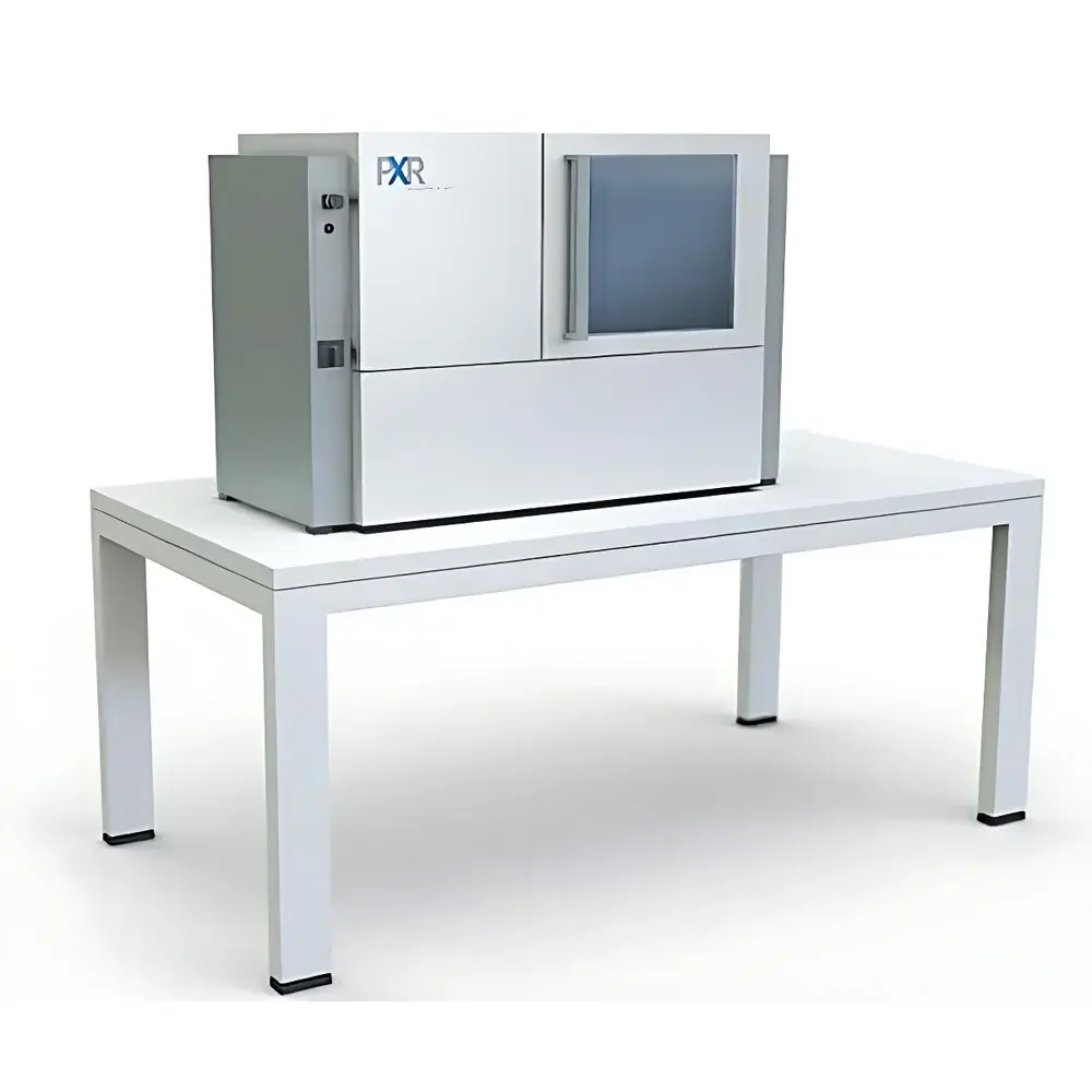
Auniontech Desktop X-ray Micro-CT System
| Brand | Auniontech |
|---|---|
| Model | micro-CT |
| X-ray Tube Voltage | Up to 200 kV |
| Detector Resolution | Up to 16 MP |
| Scan Speed | < 10 s per scan |
| Number of Projections | Up to 8-axis acquisition |
| Spatial Resolution | 0.4 µm – 5 µm |
| System Weight | 20 kg (CT-PORTABLE variant) |
| Radiation Leakage | < 1 µSv/h |
| Power Output | Up to 65 W (CT-XPRESS variant) |
Overview
The Auniontech Desktop X-ray Micro-CT System is a high-performance, benchtop-scale computed tomography platform engineered for non-destructive 3D internal structure visualization at micron- and sub-micron resolution. Based on cone-beam X-ray microtomography principles, the system employs a microfocus X-ray source (operable from 50 kV to 200 kV) coupled with a high-dynamic-range, large-area flat-panel detector to reconstruct volumetric datasets via filtered back-projection or iterative reconstruction algorithms. Unlike synchrotron-based or industrial cabinet-style CT systems, this desktop architecture delivers laboratory-grade imaging performance without requiring dedicated shielded rooms—enabled by integrated radiation containment design and real-time dose monitoring. The system supports full geometric calibration, beam hardening correction, and ring artifact suppression algorithms to ensure quantitative fidelity across diverse material densities and geometries.
Key Features
- Microfocus X-ray source with adjustable tube voltage (50–200 kV) and power output up to 65 W (CT-XPRESS configuration), enabling optimized contrast for low-Z (e.g., polymers, biological tissue) and high-Z (e.g., metals, ceramics) samples.
- Benchtop form factor: CT-PORTABLE variant weighs only 20 kg and requires no floor anchoring or structural reinforcement—ideal for shared labs, cleanrooms, or mobile QA/QC deployment.
- High-speed acquisition: Full-volume scans completed in under 10 seconds with configurable projection count (up to 8-axis scanning geometry), minimizing motion artifacts and throughput bottlenecks.
- Sub-micron spatial resolution: Achieves isotropic voxel sizes down to 0.4 µm with appropriate magnification and detector binning—validated per ISO 15732:2021 guidelines for micro-CT resolution assessment.
- Integrated radiation safety: External leakage consistently maintained below 1 µSv/h during operation, compliant with IEC 61331-1:2014 and national occupational exposure limits for Class II B X-ray equipment.
- Modular mechanical stage: Precision motorized rotation and XYZ translation stages support multi-position stitching, tilt-series acquisition, and in situ loading (optional).
Sample Compatibility & Compliance
The system accommodates specimens ranging from <1 mm³ (e.g., MEMS devices, bone biopsies) to Ø75 mm × H100 mm (CT-MINI/CT-XPRESS configurations), with density tolerance spanning air-filled foams to stainless steel alloys. Sample holders are compatible with standard petri dishes, aluminum mounts, and custom fixtures adhering to ASTM E1441-22 Annex A3 dimensional references. All firmware and acquisition protocols conform to ISO/IEC 17025:2017 requirements for measurement traceability, and raw projection data is stored in DICOM 3.0-compliant format for auditability. Optional GLP/GMP-ready software modules provide 21 CFR Part 11-compliant electronic signatures, audit trails, and user access controls—suitable for regulated environments including medical device R&D and pharmaceutical excipient characterization.
Software & Data Management
Acquisition and reconstruction are managed through Auniontech’s proprietary CT-Suite v4.x platform, which runs on Windows 10/11 64-bit workstations. The software provides real-time preview, automatic center-of-rotation alignment, phase-retrieval-assisted edge enhancement (for low-contrast interfaces), and GPU-accelerated Feldkamp-Davis-Kress (FDK) reconstruction. Export formats include NRRD, TIFF stack, STL, and VTK for downstream analysis in Avizo, Dragonfly, or MATLAB. Metadata embedding follows DICOM PS3.3 standards—including acquisition parameters, calibration coefficients, and environmental logs—to support FAIR (Findable, Accessible, Interoperable, Reusable) data principles. Raw projection archives are checksum-verified and timestamped to ensure integrity across long-term storage and cross-laboratory collaboration.
Applications
- Non-destructive evaluation (NDE): Void detection in additive-manufactured metal parts, fiber orientation mapping in CFRP composites, solder joint integrity in PCB assemblies.
- Materials science: Pore network quantification in battery electrodes, grain boundary segmentation in sintered ceramics, crack propagation tracking under thermal cycling.
- Geosciences: Digital rock physics modeling from core plug scans, fossil morphology reconstruction, soil aggregate stability analysis.
- Life sciences: Ex vivo murine organ vasculature imaging, dental enamel demineralization kinetics, plant root architecture phenotyping.
- Electronics: Wire bond lift-off detection, encapsulant delamination in IC packages, solder paste volume verification pre-reflow.
FAQ
What is the minimum achievable voxel size, and how is it verified?
The system achieves an effective isotropic voxel size of 0.4 µm under optimal geometric magnification and detector binning conditions. Resolution validation follows ISO 15732:2021 using tungsten wire phantoms and modulation transfer function (MTF) analysis.
Does the system support time-resolved (4D) scanning?
Yes—CT-XPRESS enables repeatable rapid-scan sequences (sub-10 s per volume) suitable for in situ mechanical testing or thermal expansion studies when paired with external actuators or environmental chambers.
Is remote operation supported for multi-user lab environments?
The CT-Suite platform includes secure RDP-compatible remote access mode with session logging, concurrent user role management, and TLS 1.2 encrypted data transmission.
Can raw projection data be exported for third-party reconstruction?
Yes—unprocessed TIFF-based projection stacks with full metadata headers (including source-detector geometry, exposure time, and filter configuration) are exportable for use with TomoPy, ASTRA Toolbox, or commercial alternatives.
What regulatory documentation is provided for installation in EU or US facilities?
CE marking per 2014/35/EU (Low Voltage Directive) and 2013/35/EU (EMF Directive), plus FDA product listing (as Class II diagnostic X-ray system) and RoHS 2 compliance certificates are supplied with each unit.