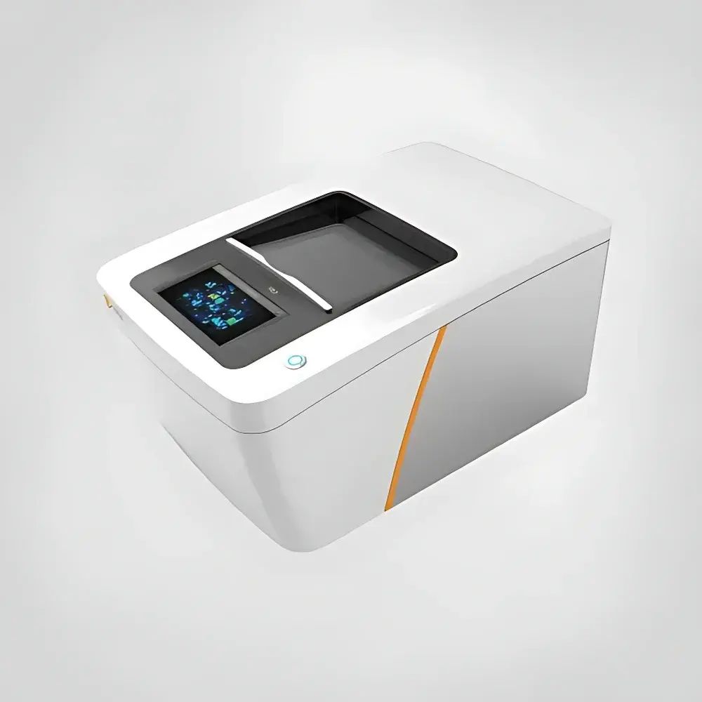
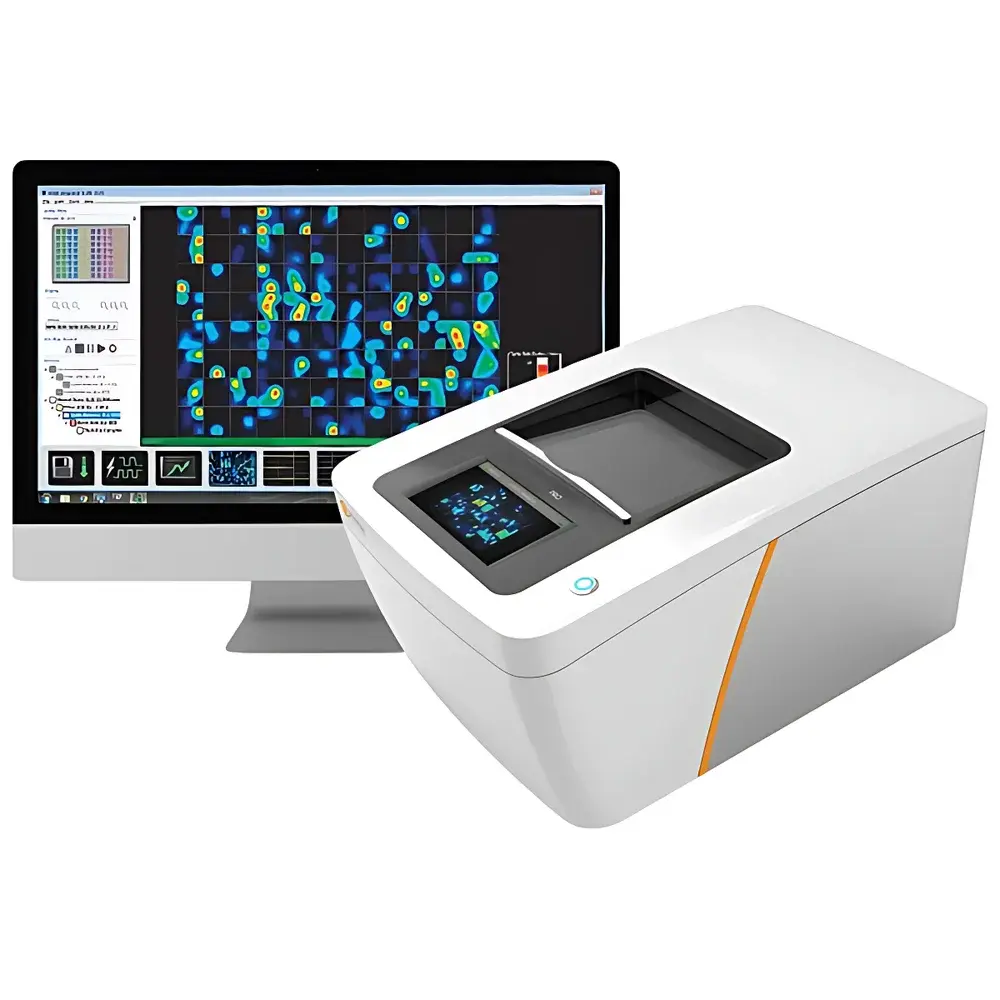
Axion BioSystems Maestro PRO/EDGE High-Throughput Microelectrode Array (MEA) System
| Brand | Axion BioSystems |
|---|---|
| Origin | USA |
| Manufacturer Type | Original Equipment Manufacturer (OEM) |
| Product Category | Imported Instrument |
| Model | Maestro PRO / EDGE |
| Throughput | Multi-well (96-well or 48-well format) |
| Application | In vitro electrophysiological recording |
| Electrode Count per Well | 384 or 768 |
| Compliance | Designed for GLP/GMP-aligned workflows, compatible with FDA 21 CFR Part 11–enabled software configurations |
Overview
The Axion BioSystems Maestro PRO/EDGE is a high-throughput, non-invasive microelectrode array (MEA) platform engineered for label-free, real-time electrophysiological monitoring of functional neural and cardiac networks in vitro. Unlike indirect surrogate assays—such as calcium imaging or transcriptional profiling—the Maestro system directly records extracellular field potentials (FPs) and action potentials (APs) with millisecond temporal resolution and spatial fidelity across hundreds of electrodes per well. Its core measurement principle is based on extracellular voltage sensing via embedded planar electrode arrays integrated into standard multiwell plates (e.g., 48- or 96-well formats), enabling simultaneous, longitudinal assessment of network-level activity from primary neurons, iPSC-derived neuronal cultures, brain slices, neurospheres, and cerebral organoids. The system supports both spontaneous and evoked activity detection, and—when paired with Axion’s Lumos optical stimulation module—enables wavelength-specific, temporally precise photostimulation (100 ms–seconds, 1–100% intensity) across up to 96 wells in parallel. This architecture provides a physiologically relevant, scalable alternative to low-throughput techniques such as manual patch clamp or single-electrode recordings.
Key Features
- High-density electrode arrays: 384 or 768 titanium-nitride microelectrodes per well, arranged in a geometric grid to resolve spatiotemporal firing patterns at cellular-network resolution.
- Real-time, long-term recording: Continuous acquisition over hours to weeks without cytotoxic dyes or genetic reporters—preserving native cell morphology, connectivity, and network integrity.
- Integrated environmental control: On-board temperature regulation (37 °C ± 0.2 °C) and CO₂/O₂ gas mixing (5% CO₂, adjustable O₂) ensure stable physiological conditions during extended experiments.
- Multi-parametric quantification: AxIS Navigator software computes primary metrics—including spike rate, burst frequency, synchrony index (cross-correlation coefficient), and oscillatory power (theta, beta, gamma bands)—as well as >25 secondary derived parameters (e.g., network burst duration, inter-burst interval variability, phase-locking value).
- Modular expandability: Seamless hardware/software integration with Lumos for optogenetic interrogation; compatibility with third-party incubators, microfluidic interfaces, and automated liquid handlers for end-to-end assay automation.
Sample Compatibility & Compliance
The Maestro platform accommodates diverse biologically relevant models: primary rodent or human cortical/hippocampal neurons; iPSC-derived excitatory/inhibitory neurons, dopaminergic or motor neurons; acute or cultured brain slices (up to 400 µm thickness); 3D neurospheres and cerebral organoids (mini-brains); co-cultures including astrocytes, microglia, or cardiomyocytes. All assays are performed under standard sterile tissue culture conditions without enzymatic dissociation or fixation. From a regulatory standpoint, the system supports audit-trail-enabled data capture (via optional 21 CFR Part 11-compliant software configuration), aligns with ISO/IEC 17025 documentation standards for method validation, and has been cited in peer-reviewed studies supporting ICH S7B and FDA guidance on nonclinical cardiovascular and neurotoxicity assessment.
Software & Data Management
AxIS Navigator serves as the central analytical engine—providing intuitive visualization tools, batch processing pipelines, and export-ready figures compliant with journal formatting requirements (TIFF, SVG, CSV, HDF5). Raw data files (.axd) are stored with embedded metadata (date/time, plate ID, protocol version, user annotation), ensuring full traceability. Advanced users may access Python and MATLAB APIs for custom algorithm development or machine learning integration (e.g., unsupervised clustering of network phenotypes). Data security protocols include role-based access control, encrypted local storage, and optional integration with enterprise LIMS or ELN systems.
Applications
- Neurodevelopmental and neurodegenerative disease modeling: Functional phenotyping of Alzheimer’s, Parkinson’s, autism spectrum disorder (ASD), epilepsy, and frontotemporal dementia using patient-derived iPSC lines.
- Neurotoxicity and safety pharmacology: Quantitative assessment of compound-induced changes in network synchrony, burst suppression, or oscillatory power—aligned with OECD TG 426 and ASTM E3232-21 guidelines.
- Drug mechanism-of-action studies: Evaluation of ion channel modulators, GPCR ligands, synaptic transmission enhancers/inhibitors, and immunomodulatory agents (e.g., IL-4, TNFα) on network excitability and homeostasis.
- Organoid maturation and quality control: Objective benchmarking of electrophysiological maturity in cerebral organoids across differentiation timepoints or bioreactor conditions.
- Neuromuscular junction modeling: Co-culture assays integrating iPSC-derived motor neurons and skeletal myotubes to assess synaptic transmission fidelity and drug-induced neuromuscular blockade.
- Translational biomarker discovery: Identification of electrophysiological endophenotypes predictive of clinical response in psychiatric or neurological indications.
FAQ
What types of cells can be recorded using the Maestro system?
Primary neurons, iPSC-derived neurons (including subtype-specific lines), brain slices, neurospheres, cerebral organoids, and cardiomyocyte monolayers or co-cultures.
Is the system compatible with optogenetics?
Yes—when integrated with the Lumos optical stimulation module, the Maestro enables wavelength-selective (450 nm, 525 nm, 590 nm, 630 nm), millisecond-precision photostimulation of opsin-expressing cells within each well.
How does Maestro differ from calcium imaging?
Calcium imaging infers electrical activity indirectly via fluorescent indicators, which suffer from slow kinetics, phototoxicity, and poor signal-to-noise for subthreshold events. Maestro measures direct extracellular voltage fluctuations with sub-millisecond resolution and no exogenous labels.
Can data be exported for statistical analysis in R or Python?
Yes—raw and processed data are exportable in standardized formats (CSV, HDF5) and accessible via documented Python/MATLAB toolkits for downstream statistical modeling, dimensionality reduction, or deep learning applications.
Does the system support GLP-compliant workflows?
With optional 21 CFR Part 11 software configuration, electronic signatures, audit trails, and secure user authentication, the Maestro platform meets key requirements for regulated preclinical safety studies.


