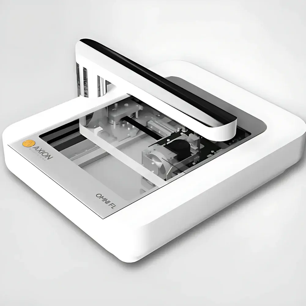
Axion BioSystems Omni BR/FL High-Content Live-Cell Imaging and Cytotoxicity Analysis System
| Brand | Axion BioSystems |
|---|---|
| Origin | Netherlands |
| Manufacturer | Axion BioSystems |
| Product Type | Imported Instrument |
| Model | Omni BR/FL |
| Temperature Control Range | 5–40 °C |
| Humidity Control Range | 20–95% RH |
| Imaging Speed | Full-plate scan in ≤10 minutes |
Overview
The Axion BioSystems Omni BR/FL is a compact, incubator-compatible high-content live-cell imaging platform engineered for label-free and fluorescence-based longitudinal monitoring of adherent mammalian cells in standard multiwell plates. Operating on the principle of automated wide-field brightfield (BR) and epifluorescence (FL) microscopy, the system captures spatially resolved morphological and functional dynamics—including confluence, membrane integrity, mitochondrial activity (via compatible dyes), and proliferation kinetics—without requiring plate removal from controlled environmental conditions. Its integrated optical architecture features an overhead LED illumination module and a motorized, high-resolution CMOS sensor mounted beneath the sample stage, enabling precise z-axis positioning and seamless tile-based image acquisition across the full 86 mm × 124 mm field of view. Designed specifically for deployment inside standard CO₂ incubators, the Omni BR/FL maintains operational stability under physiologically relevant temperature (5–40 °C) and humidity (20–95% RH) ranges, eliminating thermal and mechanical perturbations associated with external imaging workflows.
Key Features
- In-situ operation: Fully functional within standard humidified CO₂ incubators; no plate handling or environmental interruption required
- Dual-modality imaging: Simultaneous brightfield and fluorescence channels (with optional filter sets for GFP, RFP, Hoechst, etc.)
- High-throughput kinetics: Full 96-well plate scan completed in ≤10 minutes; configurable time-lapse intervals from minutes to days
- Modular software architecture: Cloud-native CytoSMART Suite with embedded algorithms for confluence quantification, scratch assay analysis, colony formation tracking, and fluorescent object counting
- Robust hardware design: Sealed electronics, corrosion-resistant housing, and passive thermal management compliant with ISO 13485-aligned manufacturing standards
- Regulatory-ready data handling: Audit trail logging, user access control, and exportable raw TIFF/OME-TIFF datasets supporting GLP/GMP-aligned workflows and FDA 21 CFR Part 11 compliance when deployed with validated IT infrastructure
Sample Compatibility & Compliance
The Omni BR/FL accommodates all optically transparent cell culture vessels with height <55 mm, including 6–384-well plates, Petri dishes, and flasks (T25–T225). Its standardized imaging area (86 mm × 124 mm) ensures consistent coverage across common microplate formats. The system complies with IEC 61000-6-2 (immunity) and IEC 61000-6-3 (emissions) for electromagnetic compatibility in laboratory environments. While not certified as a medical device, its analytical outputs align with ASTM E2925-21 (Standard Guide for Image-Based Cell Viability Assays) and ISO 20387:2018 (Biobanking — General requirements for biobank operations), supporting reproducible cytotoxicity assessment per OECD Test Guideline 492 (Reconstructed Human Cornea-like Epithelium Test Method for Eye Irritation).
Software & Data Management
All images are automatically uploaded to the secure, HIPAA-compliant CytoSMART Cloud platform via encrypted HTTPS transmission. Users access processed metrics—including normalized confluence curves, fluorescence intensity heatmaps, and migration velocity vectors—through a browser-based interface. The system supports DICOM-compliant metadata embedding and exports annotated results in CSV, PDF, and JSON formats. Third-party integration is enabled via RESTful API and open-source Python SDK (cytosmart-sdk), facilitating interoperability with MATLAB, Python-based scikit-image pipelines, and commercial platforms such as MetaMorph or Imaris. Audit logs record timestamped user actions, parameter changes, and processing events—essential for traceability in regulated toxicology studies.
Applications
- Quantitative cytotoxicity profiling: Real-time evaluation of chemotherapeutic agents (e.g., paclitaxel dose-response in PACO7/PACO43 co-cultures), including IC₅₀ derivation and kinetic delay-to-effect modeling
- Tumor spheroid viability assessment: Monitoring structural disintegration and metabolic decline in 3D models under targeted therapy or immune effector challenge
- Immunooncology assays: CAR-T or NK-cell mediated lysis kinetics tracked via membrane integrity dyes (e.g., propidium iodide) or caspase activation reporters
- Wound healing and collective migration: Automated scratch gap closure quantification with sub-pixel edge detection accuracy
- Clonogenic survival analysis: Long-term colony growth dynamics across entire plates, minimizing observer bias
- Transfection efficiency monitoring: Co-localization of fluorescent reporter expression with confluence maps to assess transduction kinetics
FAQ
How does the Omni BR/FL acquire images?
The system uses an overhead LED light source and a motorized CMOS sensor positioned beneath the sample stage. In brightfield mode, the sensor moves sequentially across the plate to capture ~7,850 individual tiles, which are stitched into a single 86 mm × 124 mm composite image. In fluorescence mode, users define region-of-interest coordinates per well to optimize signal-to-noise ratio and minimize phototoxicity.
Which image analysis modules are available?
Licensed modules include Brightfield/Fluorescence Confluence Analysis, Scratch Assay Quantification, Colony Formation Tracking, and Fluorescent Object Counting. Raw TIFF stacks can be downloaded for custom analysis in ImageJ, CellProfiler, or proprietary pipelines.
Can the Omni BR/FL operate inside a CO₂ incubator?
Yes. Its electronics, optics, and mechanics are rated for continuous operation at 5–40 °C and 20–95% RH, meeting typical incubator environmental specifications without condensation risk or thermal drift.
What vessel types are supported?
Any transparent culture vessel under 55 mm in height—including 6-, 24-, 48-, 96-, and 384-well plates, Petri dishes, and T-flasks—is compatible. Effective imaging is constrained to the central 86 mm × 124 mm area; peripheral regions may fall outside the field of view.
Is data export compatible with regulatory submissions?
Raw image data, processed metrics, and audit logs are exportable in formats suitable for inclusion in IND/CTA dossiers. When deployed with validated cloud infrastructure and documented SOPs, the workflow satisfies data integrity requirements under ALCOA+ principles and supports 21 CFR Part 11 compliance through role-based access and electronic signature implementation.


